Cleft Palate
Also known as: Congenital Oronasal Fistula — Palatoschisis
Introduction
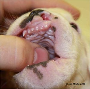
An abnormal communication between the nasal and oral cavities involving the soft palate , hard palate, premaxilla and or lip . Abnormalities arise during foetal development where there is incomplete closure of the primary palate, secondary palate or both. The primary palate develops into the premaxilla and lip. If closure is not complete this will result in a primary palate or cleft lip (harelip). The secondary palate forms the hard and soft palate and incomplete closure of either of these results in a secondary palate or cleft palate.
Possible causes may include administration of griseofulvin to queens during pregnancy, certain viral infections or ingestion of toxic plants.
Aetiology
The facial part of the dorsal surface of the skull is formed by parts of the frontal, nasal, maxillary, and incisive bones. Problems, such as a cleft palate, can arise if the fusion of these bones does not occur properly. It is also important to note that each quadrant of the skull and jaw develops independent of the other three quadrants. If there is an abnormality on one side, it does not necessarily mean the other side is also affected.
Primary cleft palate: defect between the incisive bone and maxilla. This type of cleft is often associated with a harelip or cleft lip.
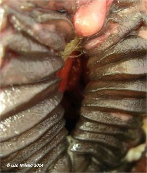
Secondary cleft palate: defect in the maxilla that may or may not extend to involve the soft palate.
Signalment
Dogs are more commonly affected than cats. In particular brachycephalic breeds are more commonly affected due to the intra-uterine growth characteristics of the skull. Other at risk dog breeds include, Boston Terriers, Pekingese, Minature Schnauzers, Beagles and Cocker Spaniels. Siamese are the most commonly affected cat breed. Present at birth but not always noticed straight away. In large animals, cleft palate has been reported in foals, calves, lambs and kids. The primary cause of this condition is often hereditary but maternal nutritional deficiencies, toxins and viral exposure have also been implemented.
Diagnosis
History and Clinical Signs
Failure to thrive with signs such as nasal discharge, nasal milk regurgitation, halitosis, gagging, coughing or sneezing during nursing, and respiratory infection due to aspiration pneumonia or rhinitis.
Diagnosis is made on physical exam. A cleft lip can be easily identified, however, a full oral examination is needed to diagnose incomplete closure of the premaxilla, hard and soft palate. In some instances anaesthesia may be required to diagnose a soft palate problem. If a primary palate problem is recognised always check for a secondary palate problem. Increased noise over lung fields may be heard if aspiration pneumonia is present. Affected animals often have other concurrent congenital abnormalities and hence a thorough physical exam should be undertaken.
Laboratory Tests
Often normal unless aspiration pneumonia is present.
Radiography
Radiographs of the skull are unnecessary however thoracic radiographs are useful to check for the presence of aspiration pneumonia.
Treatment
A large proportion of animals with primary or secondary palate defects die or are euthanased. In some cases however the defects can be managed medically until the patient is old enough for surgery.
Affected animals should gain nutritional support via a feeding tube to avoid aspiration pneumonia until surgery can be undertaken. Aspiration pneumonia should also be treated with appropriate antibiotics, expectorants and oxygen. Surgery should be delayed until the animal is 12-14 weeks old or longer if possible in order to get the best possible post surgical outcome. More mature tissue is less friable and holds suture better. Surgical correction is normally only carried out if the defect is small and owners must be warned that more severely affected animals may need multiple procedures.
Closure of Primary Clefts
Often very difficult to correct surgically, and requires planning and often multiple surgeries.
Closure of Secondary Clefts
Hard palate defects
Three procedures have been described firstly, the Lagenbeck or sliding pedicle technique whereby longitudinal strips of mucosa are released from the hard palate and slid together at the midline. Hard palate bone is exposed laterally however granulation and epithelisation occur quickly. The second technique is the Sandwich or overlapping flap technique where by a recipient bed is created by splitting the mucous membrane at the edge of the defect. A donor bed is created by releasing strips of mucosa from the opposite mucous membrane which is then sutured into place. Thirdly a combination technique can be applied where both of the above techniques are used to produce a double layer of mucosa over the cleft.
Soft Palate defects
An overlapping flap technique, flaps from the hard palate and nasopharangeal mucosa flaps can be used to repair soft palate deformities. It is important to reduce tension on suture lines, preserve the connective tissue and vascular supply and have good apposition of tissue to promote rapid healing of the wound.
Post operatively patients should be fed via a tube to prevent trauma to the surgical site and to maintain nutritional intake. Dehiscence and recurrence can occur following movement, tension and growth.
Prognosis
Historically, surgical correction of these conditions had a low success rate. More recently new surgical techniques can result in a good prognosis, however, multiple surgeries may be required and aspiration pneumonia must be treated.
| Cleft Palate Learning Resources | |
|---|---|
To reach the Vetstream content, please select |
Canis, Felis, Lapis or Equis |
 Test your knowledge using flashcard type questions |
Oral Cavity Pathology Flashcards Small Animal Soft Tissue Surgery Q&A 01 |
 Search for recent publications via CAB Abstract (CABI log in required) |
Cleft palate in Dogs publications
Cleft palate in Horses publications |
References
Fossum, T. W. et. al. (2007) Small Animal Surgery (Third Edition) Mosby Elsevier
Gilson, SD (1998) Self-Assessment Colour Review Small Animal Soft Tissue Surgery Manson
Merck & Co (2008) The Merck Veterinary Manual (Eighth Edition) Merial
| This article was expert reviewed by Lisa Milella BVSc DipEVDC MRCVS. Date reviewed: 13 August 2014 |
| Endorsed by WALTHAM®, a leading authority in companion animal nutrition and wellbeing for over 50 years and the science institute for Mars Petcare. |
Error in widget FBRecommend: unable to write file /var/www/wikivet.net/extensions/Widgets/compiled_templates/wrt661e3a15040f36_38174009 Error in widget google+: unable to write file /var/www/wikivet.net/extensions/Widgets/compiled_templates/wrt661e3a1507e5e8_31313375 Error in widget TwitterTweet: unable to write file /var/www/wikivet.net/extensions/Widgets/compiled_templates/wrt661e3a150c2b22_44362574
|
| WikiVet® Introduction - Help WikiVet - Report a Problem |
- OpenPages
- Nasal Cavity - Developmental Pathology
- Respiratory System - Developmental Pathology
- Oral Cavity - Developmental Pathology
- Oral Diseases - Cattle
- Oral Diseases - Horse
- Oral Diseases - Goat
- Oral Diseases - Pig
- Oral Diseases - Sheep
- Oral Diseases - Cat
- Oral Diseases - Dog
- Developmental Dental Conditions
- Lisa Milella reviewed
- Waltham reviewed

