File:Pericarditis-histo.jpg
Jump to navigation
Jump to search
Size of this preview: 800 × 546 pixels. Other resolutions: 320 × 218 pixels | 1,157 × 790 pixels.
Original file (1,157 × 790 pixels, file size: 160 KB, MIME type: image/jpeg)
Summary
| Description |
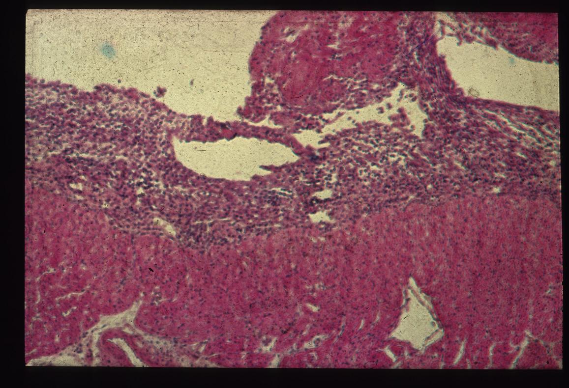
Pericarditis histology. |
|---|---|
| Date |
Unknown |
| Source |
Cambridge University |
| Author |
A. Jefferies |
| Permission (Reusing this file) |
See below |
Licensing:
| This file is licensed under the Creative Commons Attribution Non-Commercial & No Derivative Works License |
File history
Click on a date/time to view the file as it appeared at that time.
| Date/Time | Thumbnail | Dimensions | User | Comment | |
|---|---|---|---|---|---|
| current | 15:41, 29 August 2007 | | 1,157 × 790 (160 KB) | Kjr35 (talk | contribs) |
You cannot overwrite this file.
File usage
The following 2 pages use this file: