Difference between revisions of "File:Pericarditis-histo.jpg"
Jump to navigation
Jump to search
| Line 1: | Line 1: | ||
| − | The epicardium should consist of a single layer of epithelial cells in the normal heart with a similar layer lining the opposing pericardial surface. In this histological section there is an increase in cellularity of the epicardium with many inflammatory cells and pink fibrin is visible. | + | == Summary == |
| + | {{Information | ||
| + | |Description=Pericarditis histology. <br> The epicardium should consist of a single layer of epithelial cells in the normal heart with a similar layer lining the opposing pericardial surface. In this histological section there is an increase in cellularity of the epicardium with many inflammatory cells and pink fibrin is visible. | ||
| + | |Source=Cambridge University | ||
| + | |Date=Unknown | ||
| + | |Author=A. Jefferies | ||
| + | |Permission=See below | ||
| + | }} | ||
| + | |||
| + | |||
| + | == Licensing: == | ||
| + | {{cc-att-2.0}} | ||
Latest revision as of 16:07, 29 July 2010
Summary
| Description |
Pericarditis histology. |
|---|---|
| Date |
Unknown |
| Source |
Cambridge University |
| Author |
A. Jefferies |
| Permission (Reusing this file) |
See below |
Licensing:
| This file is licensed under the Creative Commons Attribution Non-Commercial & No Derivative Works License |
File history
Click on a date/time to view the file as it appeared at that time.
| Date/Time | Thumbnail | Dimensions | User | Comment | |
|---|---|---|---|---|---|
| current | 15:41, 29 August 2007 | 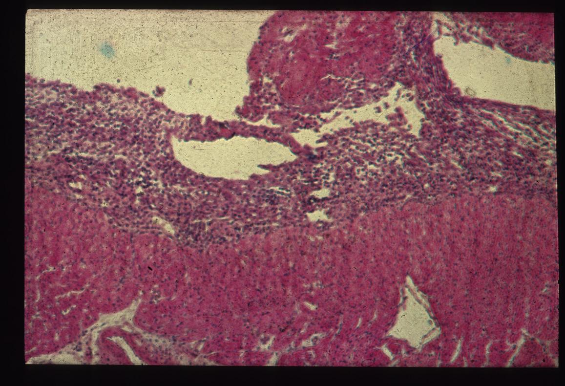 | 1,157 × 790 (160 KB) | Kjr35 (talk | contribs) |
You cannot overwrite this file.
File usage
The following 2 pages use this file: