Equine Internal Medicine Q&A 17
| This question was provided by Manson Publishing as part of the OVAL Project. See more Equine Internal Medicine questions |
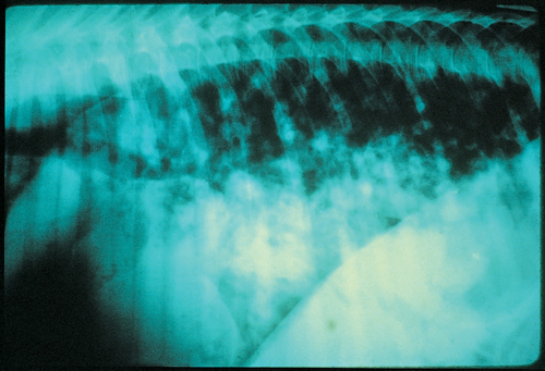
A 10-week-old foal in good body condition showed signs of acute dyspnoea.
| Question | Answer | Article | |
| Describe the radiographic findings illustrated in the image. | Lateral view of the thorax demonstrates nodular lesions in the cranioventral lung fields. These lesions, some of which are cavitary, obscure the cardiac and caudal vena cava silhouette. Tracheobronchial lymphadenopathy is suggested by the elevation of the trachea. |
[[|Link to Article]] | |
| What is the most likely aetiologic agent? | Radiographic lesions of this type seen in a 10-week-old foal are almost pathognomonic for Rhodococcus equi infection. |
Link to Article | |
| What is the source of the infection for the foal? | Rhod. equi is a Gram-positive, pleomorphic, facultative, intracellular, obligate aerobic bacteria. |
[[|Link to Article]] | |
| What is the recommended treatment? | Treatment of Rhod. equi has been difficult due to the intracellular characteristics of the organism. The combination of erythromycin and rifampin is excellent in vitro and has decreased the mortality rate associated with Rhod. equi pneumonia.
Rifampin is synergistic with erythromycin and penetrates macrophages, neutrophils and caseous material readily.
|
Link to Article | |
