Difference between revisions of "Adrenal Glands - Anatomy & Physiology"
| (31 intermediate revisions by 7 users not shown) | |||
| Line 1: | Line 1: | ||
| − | {{ | + | {{OpenPagesTop}} |
| − | + | ==Adrenal Glands== | |
| − | + | ||
| − | + | The adrenal glands are paired bodies lying cranial to the kidneys within the retroperitoneal space. The glands consist of two layers; the '''cortex''' and '''medulla'''. | |
| − | + | ||
| − | + | The '''adrenal cortex''' is red to light brown in colour and is composed of three zones. From the outer to inner, the layers are; | |
| − | |||
| − | |||
| − | + | 1. zona glomerulosa | |
| + | 2. zona fasciculata | ||
| − | + | 3. zona reticularis | |
| − | + | All these zones produce hormones derived from cholesterol, which is abundant in the cells. The '''adrenal cortex''' represents 80-90% of the adrenal gland. | |
| − | The adrenal medulla is primarily involved in the production of catecholamines; epinephrine and norepinephrine. In fetal life the adrenal medulla plays a role in the autonomic nervous system. The medulla acts as a sympathetic ganglion with the postganglionic cells lacking axons. Through sympathetic preganglionic fiber stimulation the medullary cells secrete catecholamines. The adrenal medulla represents only 10-20% of the adrenal gland. | + | The '''adrenal medulla''' is primarily involved in the production of catecholamines; epinephrine and norepinephrine. In fetal life, the adrenal medulla plays a role in the autonomic nervous system. The medulla acts as a sympathetic ganglion with the postganglionic cells lacking axons. Through sympathetic preganglionic fiber stimulation, the medullary cells secrete catecholamines. The adrenal medulla represents only 10-20% of the adrenal gland. |
[[Image:Adrenal Gland Schematic..jpg|700px|small>'''Schematic Diagram of the Adrenal Gland</small>|frameless]] | [[Image:Adrenal Gland Schematic..jpg|700px|small>'''Schematic Diagram of the Adrenal Gland</small>|frameless]] | ||
| Line 21: | Line 20: | ||
==Embryological Origin== | ==Embryological Origin== | ||
| − | |||
| − | The fetal cortex develops in the centre with the permanent cortex surrounding it. By 4 months of age the adrenal gland is fully developed. | + | The adrenal glands develop from two separate embryological tissues; the '''neural crest ectoderm''' and the '''intermediate mesoderm'''. The medulla originates from neural crest cells migrating from sympathetic ganglion. Mesodermal cells then surround the medulla. The adrenal cortex develops from the intermediate mesoderm. The fetal cortex develops in the centre, with the permanent cortex surrounding it. By 4 months of age the adrenal gland is fully developed. |
==Anatomy== | ==Anatomy== | ||
| − | Even though the adrenal glands gain | + | Even though the adrenal glands gain their name through their relationship with the kidney, they are in fact more closely related to the major vessels, including the aorta and the caudal vena cava. |
| + | |||
| + | The adrenal glands are elongated and are often asymmetrical, being moulded around the neighbouring vessels. Their size varies greatly and generally those of juveniles are larger than adults, and those of lactating or pregnant animals are larger than reproductively inactive animals. A medium-sized dog's adrenals will on average measure 2.5 x 1 x 0.5cm. The adrenal glands are firm, and their capsule is easily fractured on flexion. The cortex on appearance is yellow and radially striated, whilst the medulla is darker with a more uniform appearance. The '''zona glomerulosa''' is narrow and the cells are in a whorled pattern. The '''zona fasiculata''' is wide and the cells lie in columns and the '''zona reticularis''' is more randomly organised. | ||
| − | + | ==Vascular Supply== | |
| − | + | The oxygenated supply is from various branches of the following neighbouring trunks; aorta, renal artery, lumbar artery, phrenicoabdominal artery and the cranial mesenteric arteries. After perfusion of the gland, the blood pools in a central vein and then exits the gland through the hilus. This then joins up with the caudal vena cava or one of its tributaries. | |
| − | The | + | ==Function== |
| + | |||
| + | ===Adrenal Cortex=== | ||
| + | The function of the cortex is to produce '''adrenocorticoid hormones'''. The three zones each make a specific type of hormone: | ||
| − | + | The Zona Glomerulosa secretes '''mineralocorticoids''', | |
| − | The | + | The Zona Fasciculata secretes '''glucocorticoids''', |
| − | + | The Zona Reticularis secretes sex steroids or '''androgens'''. | |
| − | + | The hormones are formed from the esterified cholesterol that is stored in intracellular lipid droplets. Below is the steps of how the adrenocorticoid hormones are made from cholesterol. | |
| − | |||
| − | |||
| − | |||
| − | |||
| − | |||
| − | |||
1) The P450scc enzyme converts cholesterol to pregnenolone by the removal of a side chain. This is regulated by ACTH and is the rate determining step of adrenocorticoid hormone production. This occurs in the mitochondria. | 1) The P450scc enzyme converts cholesterol to pregnenolone by the removal of a side chain. This is regulated by ACTH and is the rate determining step of adrenocorticoid hormone production. This occurs in the mitochondria. | ||
| + | |||
2) Hydroxylation reactions occur in the mitochondria and endoplasmic reticulum converting pregnenolone into specific hormones. The location within the cortex determines the hormones produced. | 2) Hydroxylation reactions occur in the mitochondria and endoplasmic reticulum converting pregnenolone into specific hormones. The location within the cortex determines the hormones produced. | ||
| − | + | ====Mineralocorticoids==== | |
| − | These hormones regulate the | + | These hormones regulate the metabolism of inorganic ions, such as sodium, potassium and chloride. Several hormones are produced but the most important is '''aldosterone'''. [[Aldosterone]] is formed in the zona glomerulosa by the conversion of pregnenolone to cortisol and then to corticosterone, before finally becoming aldosterone. It circulates by binding to albumin and cortisol-binding globulin. It's plasma half-life is 20 minutes. It is eventually inactivated in the liver, where it binds to glucuronic acid and is excreted in bile and urine. |
| − | + | ||
| + | '''Regulation and action''' | ||
| + | |||
| + | It's main regulators of synthesis and secretion are; '''Renin''' and extracellular concentrations of potassium ions. Its main action is upon renal regulation and electrolyte and fluid balance. These are fully explained within the '''[[Renin Angiotensin Aldosterone System|renin-angiotensin]] and [[Aldosterone|aldosterone system]] pages. | ||
| + | |||
| + | ====Glucocorticoids==== | ||
| + | These hormones are important in regulating glucose metabolism; the most important of them is '''cortisol'''. This is formed within the zona fasciculata by the conversion of pregnenolone directly to cortisol. It is transported by cortisol-binding globulin and has a half-life of 90 minutes. | ||
| + | |||
| + | '''Regulation of Synthesis and Secretion''' | ||
| + | |||
| + | This is regulated by the hypothalamus-pituitary adrenal axis as follows: | ||
| + | [[Image:ACTH axis.jpg|right|thumb|205px|<small><center>'''Schematic Diagram of the Hypothalamic Pituitary Adrenal Axis, Copyright John Tulloch</center></small>]] | ||
| + | |||
| + | An environmental, or internal stimuli (such as stress or light regulating the circadian rhythm) causes the hypothalamus to release ACTH-releasing hormone ('''ACTH-RH'''). ACTH-RH flows to the pituitary gland, stimulating the production of adrenocorticotrophic hormone ('''ACTH'''). ACTH binds to receptors on the cells of the adrenal cortex, thus activating adenyl cyclase. This leads to increased cAMP levels, and the mobilization of more cholesterol and so the production of more cortisol. This is the rate limiting step and is determined by the P-450-enzyme. Cortisol then has a negative feedback effect on ACTH-RH and ACTH secretion. | ||
| + | |||
| + | '''Action''' | ||
| + | |||
| + | These are essential hormones and have multiple effects around the body. Cortisol has a '''permissive action''', i.e. without cortisol certain chemical reactions within cells are unable to occur, for example the production of epinephrine and glucagon. Cortisol is a '''stress hormone''', in times of stress it is released and enhances the effect of norepinephrine on blood pressure. It is also essential for the day to day maintenance of normal blood pressure and will also stimulate gluconeogenesis and inhibit certain tissues utilization of glucose. Cortisol stimulates the degradation of fats and proteins and inhibits DNA synthesis in some tissues. This along with degradation of protein, results in a '''growth-inhibiting effect''' in times of stress. It can also have '''anti-inflammatory''' effects. It inhibits the formation of prostaglandins and cytokines and reduces the ability of white blood cells being transported to an injured area. It helps keep in check inflammatory processes that could potentially cause tissue damage. Cortisol is also '''immunosuppressive''', by reducing the lymphocyte numbers in circulation and reducing antibody production. | ||
| + | |||
| + | ====Androgens==== | ||
| + | These hormones function as male sex hormones. The most important of these is '''testosterone'''. This is produced by the conversion of pregnenolone to dehydroepiandrosterone, then to androstenedione and finally to testosterone. This occurs in the zona reticularis. They bind in the blood to sex hormone-binding globulin (SHBG) and albumin. Most adrenal androgens have little physiological action until they are converted to testosterone. They have little importance within the male, however, in women adrenal androgens, which are converted to oestrogen in adipose tissue, is the most important source of oestrogen after the menopause. | ||
| + | |||
| + | ===Adrenal Medulla=== | ||
| + | |||
| + | The adrenal medulla converts the amino acid to '''catecholamines'''. This group of hormones contains '''epinephrine''' and '''norepinephrine'''. All of the bodies circulating epinephrine is produced by the adrenal medulla, where as norepinephrine comes from both the medulla and postganglionic sympathetic neurons. | ||
| + | |||
| + | '''Synthesis, secretion and metabolism''' | ||
| + | |||
| + | Tyrosine is firstly converted to dihydroxyphenylalanine by tyrosine hydroxylase. This is the rate-limiting step. Dihydroxyphenylalanine is then converted into dopamine, then into norepinephrine and finally into epinephrine. They are stored in secretory vesicles and are released by exocytosis. They are metabolised in the liver and kidney and their half-lives are only 1-3 minutes. Some unmetabolised catecholamines are excreted in the urine. | ||
| + | |||
| + | '''Regulation of synthesis and secretion''' | ||
| + | |||
| + | They are released in response to stress by the firing of preganglionic sympathetic nerve fibres in the adrenal medulla. This causes release of acetylcholine, thus depolarizing the cell membrane. An action potential is generated and an influx of calcium ions results in the exocytosis of the catecholamine vesicles. | ||
| + | |||
| + | '''Adrenergic Receptors''' | ||
| + | |||
| + | The catecholamines bind to two types of receptor proteins in cell membranes to elicit a response; these are alpha and beta receptors. The following is a table of where the receptors are located in the body and their effects when stimulated. | ||
| − | '' | + | <TABLE CELLPADDING="3" CELLSPACING="3" > |
| + | <TR> | ||
| + | <TD BGCOLOR="#C1f0f6"><center>'''Target Tissue'''</center></TD> | ||
| + | <TD BGCOLOR="#C1f0f6"><center>'''Receptor Type'''</center></TD> | ||
| + | <TD BGCOLOR="#C1f0f6"><center>'''Responses'''</center></TD> | ||
| + | </TR> | ||
| + | <TR> | ||
| + | <TD><center>Eye</center></TD> | ||
| + | <TD><center>alpha-1</center></TD> | ||
| + | <TD><center>radial muscle contraction</center></TD> | ||
| + | </TR> | ||
| + | <TR> | ||
| + | <TD BGCOLOR="#C1f0f6"><center>Pancreas</center></TD> | ||
| + | <TD BGCOLOR="#C1f0f6"><center>alpha-2</center></TD> | ||
| + | <TD BGCOLOR="#C1f0f6"><center>decreased insulin secretion</center></TD> | ||
| + | </TR> | ||
| + | <TR> | ||
| + | <TD><center>Cardiovascular system</center></TD> | ||
| + | <TD><center>alpha-2</center></TD> | ||
| + | <TD><center>vasoconstriction</center></TD> | ||
| + | </TR> | ||
| + | <TR> | ||
| + | <TD BGCOLOR="#C1f0f6"><center>urinary bladder</center></TD> | ||
| + | <TD BGCOLOR="#C1f0f6"><center>alpha-2</center></TD> | ||
| + | <TD BGCOLOR="#C1f0f6"><center>sphincter contraction</center></TD> | ||
| + | <TR> | ||
| + | <TD><center>uterus</center></TD> | ||
| + | <TD><center>alpha-2</center></TD> | ||
| + | <TD><center>contraction</center></TD> | ||
| + | </TR> | ||
| + | <TR> | ||
| + | <TD BGCOLOR="#C1f0f6"><center>male sex organs</center></TD> | ||
| + | <TD BGCOLOR="#C1f0f6"><center>alpha-2</center></TD> | ||
| + | <TD BGCOLOR="#C1f0f6"><center>ejaculation, detumescence</center></TD> | ||
| + | <TR> | ||
| + | <TD><center>central nervous system</center></TD> | ||
| + | <TD><center>alpha-2</center></TD> | ||
| + | <TD><center>stimulation</center></TD> | ||
| + | </TR> | ||
| + | <TR> | ||
| + | <TD BGCOLOR="#C1f0f6"><center>skin</center></TD> | ||
| + | <TD BGCOLOR="#C1f0f6"><center>alpha-2</center></TD> | ||
| + | <TD BGCOLOR="#C1f0f6"><center>piloerection, sweat production</center></TD> | ||
| + | <TR> | ||
| + | <TD><center>cardiovascular system</center></TD> | ||
| + | <TD><center>beta-1</center></TD> | ||
| + | <TD><center>increased heart rate, increased contractility, increased conduction velocity</center></TD> | ||
| + | </TR> | ||
| + | <TR> | ||
| + | <TD BGCOLOR="#C1f0f6"><center>renin secretion</center></TD> | ||
| + | <TD BGCOLOR="#C1f0f6"><center>beta-1</center></TD> | ||
| + | <TD BGCOLOR="#C1f0f6"><center>stimulation</center></TD> | ||
| + | <TR> | ||
| + | <TD><center>liver</center></TD> | ||
| + | <TD><center>beta-2</center></TD> | ||
| + | <TD><center>glycogenolysis, lipolysis, gluconeogenesis</center></TD> | ||
| + | </TR> | ||
| + | <TR> | ||
| + | <TD BGCOLOR="#C1f0f6"><center>adipose tissue</center></TD> | ||
| + | <TD BGCOLOR="#C1f0f6"><center>beta-2</center></TD> | ||
| + | <TD BGCOLOR="#C1f0f6"><center>lipolysis</center></TD> | ||
| + | <TR> | ||
| + | <TD><center>skeletal muscle</center></TD> | ||
| + | <TD><center>beta-2</center></TD> | ||
| + | <TD><center>glycogenolysis</center></TD> | ||
| + | </TR> | ||
| + | <TR> | ||
| + | <TD BGCOLOR="#C1f0f6"><center>pancreas</center></TD> | ||
| + | <TD BGCOLOR="#C1f0f6"><center>beta-2</center></TD> | ||
| + | <TD BGCOLOR="#C1f0f6"><center>increased insulin secretion</center></TD> | ||
| + | <TR> | ||
| + | <TD><center>cardiovascular system</center></TD> | ||
| + | <TD><center>beta-2</center></TD> | ||
| + | <TD><center>vasodilation in skeletal muscle arterioles, coronary arteries and all veins</center></TD> | ||
| + | </TR> | ||
| + | <TR> | ||
| + | <TD BGCOLOR="#C1f0f6"><center>bronchial muscles</center></TD> | ||
| + | <TD BGCOLOR="#C1f0f6"><center>beta-2</center></TD> | ||
| + | <TD BGCOLOR="#C1f0f6"><center>relaxation</center></TD> | ||
| + | <TR> | ||
| + | <TD><center>gastrointestinal tract</center></TD> | ||
| + | <TD><center>beta-2</center></TD> | ||
| + | <TD><center>relaxation</center></TD> | ||
| + | </TR> | ||
| + | <TR> | ||
| + | <TD BGCOLOR="#C1f0f6"><center>urinary bladder</center></TD> | ||
| + | <TD BGCOLOR="#C1f0f6"><center>beta-2</center></TD> | ||
| + | <TD BGCOLOR="#C1f0f6"><center>detrusor relaxation</center></TD> | ||
| + | <TR> | ||
| + | <TD><center>uterus</center></TD> | ||
| + | <TD><center>beta-2</center></TD> | ||
| + | <TD><center>relaxation</center></TD> | ||
| + | </TR> | ||
| + | <TR> | ||
| + | <TD BGCOLOR="#C1f0f6"><center>male sex organs</center></TD> | ||
| + | <TD BGCOLOR="#C1f0f6"><center>beta-2</center></TD> | ||
| + | <TD BGCOLOR="#C1f0f6"><center>erection?</center></TD> | ||
| + | <TR> | ||
| + | <TD><center>eye</center></TD> | ||
| + | <TD><center>beta-2</center></TD> | ||
| + | <TD><center>ciliary muscle relaxation</center></TD> | ||
| + | </TR></TABLE> | ||
| + | |||
| + | ==Histology== | ||
| − | |||
| − | |||
| − | |||
| − | |||
| − | |||
<gallery> | <gallery> | ||
| + | Image:Histology of the Adrenal Glands..jpg|<small>'''Histological section of the Adrenal Gland, from the [[Endocrine Histology resource|Histology of the Endocrine system tutorial]]</small> | ||
| + | Image:Histology of the Adrenal Glands showing zones..jpg|<small>'''Histological section of the Adrenal Gland Cortical zones, from the [[Endocrine Histology resource|Histology of the Endocrine system tutorial]]</small> | ||
| + | Image:Histology of the Adrenal Glands Medulla..jpg|<small>'''Histological section of the Adrenal Gland showing Medulla and Zona Reticularis, from the [[Endocrine Histology resource|Histology of the Endocrine system tutorial]]</small> | ||
| + | </gallery> | ||
| + | |||
| + | ==Functional anatomy== | ||
| + | [[Image:Normal adrenal cortex.jpg|right|thumb|125px|<small><center>'''Normal Adrenal Cortex'''. Courtesy of A. Jefferies</center></small>]] | ||
| + | The adrenal glands are essential to life. | ||
| + | They are closely applied to the cranial poles of each kidney. Consists of a cortex and medulla, each having different embryological origin. In some lower animals the two components actually exist as separate endcrine glands. | ||
| + | *'''Cortex''': Secretes a variety of steroid hormones derived from ''cholesterol''. | ||
| + | **''Zona glomerulosa'': Secretes '''Aldosterone'''. Concerned with electrolyte and fluid homeostasis. | ||
| + | **''Zona Fasciculata'': Secretes '''Glucocorticoids'''. Controls metabolism of lipid, protein and carbohydrate. | ||
| + | **''Zona Reticularis'': Produces '''Sex hormones'''. Supplements gonadal sex hormone secretion. | ||
| + | *'''Medulla''': Embryologically similar origin to the sympathetic nervous system. Secretes ''catecholamine hormones'' E.g. Adrenaline, noradrenaline.<br> | ||
| + | Secretion from the medulla is directly controlled by the sympathetic nervous | ||
| + | system, allowing a rapid response. In contrast, the secretions from the cortex are controlled by the hormones ACTH and other circulating hormones. | ||
| + | |||
| + | As with any endocrine gland there are two main types of pathology: | ||
| + | *Insufficiency leading to [[Hypoadrenocorticism|'''Addisons disease''']]. | ||
| + | *Excess leading to [[Hyperadrenocorticism|'''Cushings disease''']]. | ||
| + | |||
| + | |||
| + | |||
| + | {{Template:Learning | ||
| + | |powerpoints = [[Endocrine Histology resource|Histology of the Endocrine system]] | ||
| + | |OVAM = [http://www.onlineveterinaryanatomy.net/content/ovine-adrenal-gland-histology Ovine Adrenal Gland Histology 1]<br>[http://www.onlineveterinaryanatomy.net/content/ovine-adrenal-gland-histology-zona-fasiculate-and-zona-glomerulosa Ovine Adrenal Gland Histology 2] | ||
| + | |Vetstream = [https://www.vetstream.com/canis/search?s=adrenals Adrenal diseases] | ||
| + | }} | ||
| + | |||
| + | {{OpenPages}} | ||
| + | [[Category:Endocrine System - Anatomy & Physiology]] | ||
| + | [[Category:A&P Done]] | ||
Revision as of 20:36, 17 May 2016
Adrenal Glands
The adrenal glands are paired bodies lying cranial to the kidneys within the retroperitoneal space. The glands consist of two layers; the cortex and medulla.
The adrenal cortex is red to light brown in colour and is composed of three zones. From the outer to inner, the layers are;
1. zona glomerulosa
2. zona fasciculata
3. zona reticularis
All these zones produce hormones derived from cholesterol, which is abundant in the cells. The adrenal cortex represents 80-90% of the adrenal gland.
The adrenal medulla is primarily involved in the production of catecholamines; epinephrine and norepinephrine. In fetal life, the adrenal medulla plays a role in the autonomic nervous system. The medulla acts as a sympathetic ganglion with the postganglionic cells lacking axons. Through sympathetic preganglionic fiber stimulation, the medullary cells secrete catecholamines. The adrenal medulla represents only 10-20% of the adrenal gland.

Embryological Origin
The adrenal glands develop from two separate embryological tissues; the neural crest ectoderm and the intermediate mesoderm. The medulla originates from neural crest cells migrating from sympathetic ganglion. Mesodermal cells then surround the medulla. The adrenal cortex develops from the intermediate mesoderm. The fetal cortex develops in the centre, with the permanent cortex surrounding it. By 4 months of age the adrenal gland is fully developed.
Anatomy
Even though the adrenal glands gain their name through their relationship with the kidney, they are in fact more closely related to the major vessels, including the aorta and the caudal vena cava.
The adrenal glands are elongated and are often asymmetrical, being moulded around the neighbouring vessels. Their size varies greatly and generally those of juveniles are larger than adults, and those of lactating or pregnant animals are larger than reproductively inactive animals. A medium-sized dog's adrenals will on average measure 2.5 x 1 x 0.5cm. The adrenal glands are firm, and their capsule is easily fractured on flexion. The cortex on appearance is yellow and radially striated, whilst the medulla is darker with a more uniform appearance. The zona glomerulosa is narrow and the cells are in a whorled pattern. The zona fasiculata is wide and the cells lie in columns and the zona reticularis is more randomly organised.
Vascular Supply
The oxygenated supply is from various branches of the following neighbouring trunks; aorta, renal artery, lumbar artery, phrenicoabdominal artery and the cranial mesenteric arteries. After perfusion of the gland, the blood pools in a central vein and then exits the gland through the hilus. This then joins up with the caudal vena cava or one of its tributaries.
Function
Adrenal Cortex
The function of the cortex is to produce adrenocorticoid hormones. The three zones each make a specific type of hormone:
The Zona Glomerulosa secretes mineralocorticoids,
The Zona Fasciculata secretes glucocorticoids,
The Zona Reticularis secretes sex steroids or androgens.
The hormones are formed from the esterified cholesterol that is stored in intracellular lipid droplets. Below is the steps of how the adrenocorticoid hormones are made from cholesterol.
1) The P450scc enzyme converts cholesterol to pregnenolone by the removal of a side chain. This is regulated by ACTH and is the rate determining step of adrenocorticoid hormone production. This occurs in the mitochondria.
2) Hydroxylation reactions occur in the mitochondria and endoplasmic reticulum converting pregnenolone into specific hormones. The location within the cortex determines the hormones produced.
Mineralocorticoids
These hormones regulate the metabolism of inorganic ions, such as sodium, potassium and chloride. Several hormones are produced but the most important is aldosterone. Aldosterone is formed in the zona glomerulosa by the conversion of pregnenolone to cortisol and then to corticosterone, before finally becoming aldosterone. It circulates by binding to albumin and cortisol-binding globulin. It's plasma half-life is 20 minutes. It is eventually inactivated in the liver, where it binds to glucuronic acid and is excreted in bile and urine.
Regulation and action
It's main regulators of synthesis and secretion are; Renin and extracellular concentrations of potassium ions. Its main action is upon renal regulation and electrolyte and fluid balance. These are fully explained within the renin-angiotensin and aldosterone system pages.
Glucocorticoids
These hormones are important in regulating glucose metabolism; the most important of them is cortisol. This is formed within the zona fasciculata by the conversion of pregnenolone directly to cortisol. It is transported by cortisol-binding globulin and has a half-life of 90 minutes.
Regulation of Synthesis and Secretion
This is regulated by the hypothalamus-pituitary adrenal axis as follows:

An environmental, or internal stimuli (such as stress or light regulating the circadian rhythm) causes the hypothalamus to release ACTH-releasing hormone (ACTH-RH). ACTH-RH flows to the pituitary gland, stimulating the production of adrenocorticotrophic hormone (ACTH). ACTH binds to receptors on the cells of the adrenal cortex, thus activating adenyl cyclase. This leads to increased cAMP levels, and the mobilization of more cholesterol and so the production of more cortisol. This is the rate limiting step and is determined by the P-450-enzyme. Cortisol then has a negative feedback effect on ACTH-RH and ACTH secretion.
Action
These are essential hormones and have multiple effects around the body. Cortisol has a permissive action, i.e. without cortisol certain chemical reactions within cells are unable to occur, for example the production of epinephrine and glucagon. Cortisol is a stress hormone, in times of stress it is released and enhances the effect of norepinephrine on blood pressure. It is also essential for the day to day maintenance of normal blood pressure and will also stimulate gluconeogenesis and inhibit certain tissues utilization of glucose. Cortisol stimulates the degradation of fats and proteins and inhibits DNA synthesis in some tissues. This along with degradation of protein, results in a growth-inhibiting effect in times of stress. It can also have anti-inflammatory effects. It inhibits the formation of prostaglandins and cytokines and reduces the ability of white blood cells being transported to an injured area. It helps keep in check inflammatory processes that could potentially cause tissue damage. Cortisol is also immunosuppressive, by reducing the lymphocyte numbers in circulation and reducing antibody production.
Androgens
These hormones function as male sex hormones. The most important of these is testosterone. This is produced by the conversion of pregnenolone to dehydroepiandrosterone, then to androstenedione and finally to testosterone. This occurs in the zona reticularis. They bind in the blood to sex hormone-binding globulin (SHBG) and albumin. Most adrenal androgens have little physiological action until they are converted to testosterone. They have little importance within the male, however, in women adrenal androgens, which are converted to oestrogen in adipose tissue, is the most important source of oestrogen after the menopause.
Adrenal Medulla
The adrenal medulla converts the amino acid to catecholamines. This group of hormones contains epinephrine and norepinephrine. All of the bodies circulating epinephrine is produced by the adrenal medulla, where as norepinephrine comes from both the medulla and postganglionic sympathetic neurons.
Synthesis, secretion and metabolism
Tyrosine is firstly converted to dihydroxyphenylalanine by tyrosine hydroxylase. This is the rate-limiting step. Dihydroxyphenylalanine is then converted into dopamine, then into norepinephrine and finally into epinephrine. They are stored in secretory vesicles and are released by exocytosis. They are metabolised in the liver and kidney and their half-lives are only 1-3 minutes. Some unmetabolised catecholamines are excreted in the urine.
Regulation of synthesis and secretion
They are released in response to stress by the firing of preganglionic sympathetic nerve fibres in the adrenal medulla. This causes release of acetylcholine, thus depolarizing the cell membrane. An action potential is generated and an influx of calcium ions results in the exocytosis of the catecholamine vesicles.
Adrenergic Receptors
The catecholamines bind to two types of receptor proteins in cell membranes to elicit a response; these are alpha and beta receptors. The following is a table of where the receptors are located in the body and their effects when stimulated.
Histology
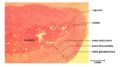
Histological section of the Adrenal Gland, from the Histology of the Endocrine system tutorial
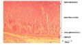
Histological section of the Adrenal Gland Cortical zones, from the Histology of the Endocrine system tutorial
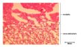
Histological section of the Adrenal Gland showing Medulla and Zona Reticularis, from the Histology of the Endocrine system tutorial


Functional anatomy

The adrenal glands are essential to life. They are closely applied to the cranial poles of each kidney. Consists of a cortex and medulla, each having different embryological origin. In some lower animals the two components actually exist as separate endcrine glands.
- Cortex: Secretes a variety of steroid hormones derived from cholesterol.
- Zona glomerulosa: Secretes Aldosterone. Concerned with electrolyte and fluid homeostasis.
- Zona Fasciculata: Secretes Glucocorticoids. Controls metabolism of lipid, protein and carbohydrate.
- Zona Reticularis: Produces Sex hormones. Supplements gonadal sex hormone secretion.
- Medulla: Embryologically similar origin to the sympathetic nervous system. Secretes catecholamine hormones E.g. Adrenaline, noradrenaline.
Secretion from the medulla is directly controlled by the sympathetic nervous system, allowing a rapid response. In contrast, the secretions from the cortex are controlled by the hormones ACTH and other circulating hormones.
As with any endocrine gland there are two main types of pathology:
- Insufficiency leading to Addisons disease.
- Excess leading to Cushings disease.
| Adrenal Glands - Anatomy & Physiology Learning Resources | |
|---|---|
To reach the Vetstream content, please select |
Canis, Felis, Lapis or Equis |
 Selection of relevant PowerPoint tutorials |
Histology of the Endocrine system |
Anatomy Museum Resources |
Ovine Adrenal Gland Histology 1 Ovine Adrenal Gland Histology 2 |
Error in widget FBRecommend: unable to write file /var/www/wikivet.net/extensions/Widgets/compiled_templates/wrt6621982542d210_58865840 Error in widget google+: unable to write file /var/www/wikivet.net/extensions/Widgets/compiled_templates/wrt662198254711d8_56760609 Error in widget TwitterTweet: unable to write file /var/www/wikivet.net/extensions/Widgets/compiled_templates/wrt662198254a8ac6_63954453
|
| WikiVet® Introduction - Help WikiVet - Report a Problem |