Adrenal Glands - Anatomy & Physiology
|
|
Adrenal Glands
The adrenal glands are paired bodies lying cranial to the kidneys within the retroperitoneal space. The glands consist of two layers; the cortex and medulla.
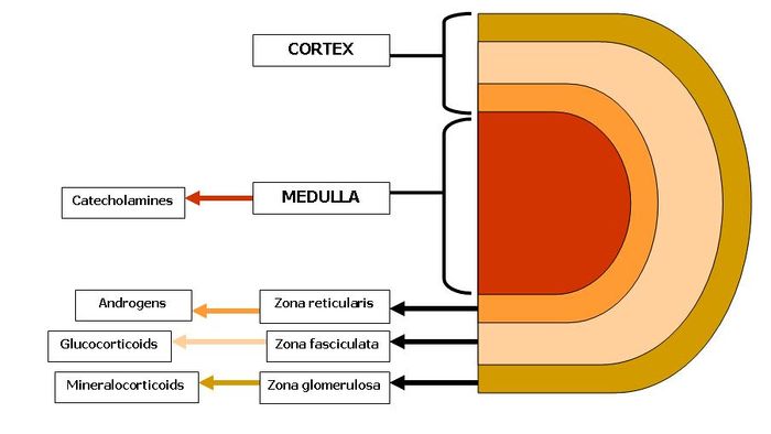
The adrenal cortex is red - light brown in colour and is comprised of three zones. These zones all produce hormones derived from cholesterol which is abundant in the cells. From outer to inner the layers are zona glomerulosa, zona fasciculata and zona reticularis. The adrenal cortex represents 80-90% of the adrenal gland.
The adrenal medulla is primarily involved in the production of catecholamines; epinephrine and norepinephrine. In fetal life the adrenal medulla plays a role in the autonomic nervous system. The medulla acts as a sympathetic ganglion with the postganglionic cells lacking axons. Through sympathetic preganglionic fiber stimulation the medullary cells secrete catecholamines. The adrenal medulla represents only 10-20% of the adrenal gland.
Embryological Origin
The adrenal glands develop from two separate embryological tissues; the neural crest ectoderm and the intermediate mesoderm. The adrenal cortex develops from the intermediate mesoderm. The medulla originates from neural crest cells migrating from sympathetic ganglion. Mesodermal cells then surround the medulla.
The fetal cortex develops in the centre with the permanent cortex surrounding it. By 4 months of age the adrenal gland is fully developed.
Anatomy
Even though the adrenal glands gain through name through their relationship with the kidney they are in fact more closely related to the major vessels. They are closely connected to the aorta and caudal vena cava.
They are elongated and are often asymetrical being moulderd around the neighbouring vessels. Their size varies a lot and generally those of juveniles are larger than adults, and those of lactatcing or pregnant animals are larger than reproductively inactive animals. A medium-sized dog's adrenals will on average measure 2.5x1x0.5cm.
They are firm and their capsule is easily fractured on flexion. The cortex on appearance is yellow and radially striated whilst the medulla is darker with a more uniform appearance.
The zona glomerulosa is narrow and the cells are in a whorled pattern. The zona fasiculata is wide and the cells lie in columns. The zona reticularis are more randomly organised.
Vascular Supply
The oxygenated supply is from various branches of the following neighbouring trunks; aorta, renal artery, lumbar artery, phrenicoabdominal artery and the cranial mesenteric arteries.
After perfusion of the gland the blood pools in a central vein and then exit the gland through the hilus. This then joins up with the caudal vena cava or one of it's tributaries.
Function
Adrenal Cortex
- The Zona Glomerulosa secretes mineralocorticoids
- The Zona Fasciculata secretes glucocorticoids
- The Zona Reticularis secretes sex steroids
The Synthesis and Metabolism of Adrenal Corticoids
The synthesis of these corticoids is through the involvement of the steroid biosynthesis pathway. The steps of which are outlined below: 1) An enzyme cleaves the carbon side chain of cholesterol esters, forming the C21 steroid; Pergnenolone 2) Hydroxylations at C-21, ensures that these molecules then don't enter the progesterone pathway.
Histology of the Adrenal Glands
<gallery> Image:Histology of the Adrenal Glands..jpg|Histological section of the Adrenal Gland Image:Histology of the Adrenal Glands showing zones..jpg|Histological section of the Adrenal Gland Cortical zones Image:Histology of the Adrenal Glands Medulla..jpg|Histological section of the Adrenal Gland showing Medulla and Zona Reticularis <gallery>
