Avian Intestines - Anatomy & Physiology
Jump to navigation
Jump to search
|
|
Structure
- The inestines occupy the caudal part of the body
- Contacts the reproductive organs and gizzard
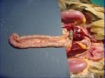
- The small intestine is long and relatively uniform in shape and size
- The duodenum passes caudally over the gizzard then loops back towards the stomach where it joins the jejunum
- Jejunum
- Loose coils around the mesentery
- Thin walls so content appears green
- Suspended from the dorsal wall of the abdomen by the mesentery
- Ileum
- Begins opposite the apices of the caeca or at the vitelline diverticula
- Suspended from the dorsal wall of the abdomen by the mesentery
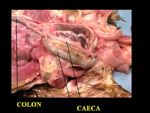
- A short colon
- The colon lies ventral to the synsacrum and opens into the cloaca
- Runs ventral to the vertebrae
- Terminates in the coprodeum
- Amino acids and glucose can be absorbed
- 2 caeca from the ileocaecal junction run with the ileum caudally
- Blind sacs about 16-18cm long
- Extend towards the liver then fold back on themselves
- Mesentery runs between the caeca then on towards the ileum
- Often contain dark coloured material
- 3 parts of each caeca
- Bacterial breakdown of cellulose occurs
- Antiperistaltic movements transport chyme
- Caeca emptied a few times per day
- Unlike mammals, there are no lacteals in the epithelium
Vitelline Diverticula
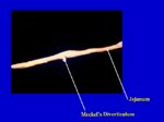
- Small outgrowth on the jejunum
- Former connection will yolk sac
- Also called Meckel's diverticulum
Function
- See small intestine
Vasculature
- See small intestine
Innervation
- See small intestine
Lymphatics
- Patches of lymphoid nodules are present in Peyer's Patches
- Most abundant in the duodenum
- No mesenteric lymph nodes
Histology
- Caeca
- Serous coat has nerve plexuses
- Columnar epithelium and goblet cells
- Smooth muscle in folds at base
- Caecal sphincter at proximal part containing a lot of lymphoid tissue (caecal tonsil)
- Middle section has thin walls and appears green
- The bulbous blind ends have thicker walls
- See small intestine
Species Differences
- Duck and goose have several loops of 'U' shaped jejunum
- Pigeons have a circular mass of jejunum with inner and outer turns
- Long caeca in turkey and chicken
- Pigeons and song birds have short caeca
- Parrots do not have caeca
- The dorsal and ventral lobes of the pancreas are connected dorsally in poultry
Links
Avian Alimentary Tract Flashcards


