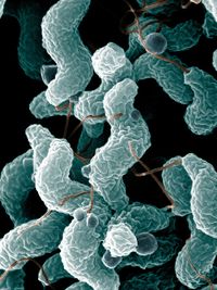
Campylobacteriosis - Cattle
Introduction
Gastrointestinal campylobacteriosis is caused by Campylobacter jejuni or Campylobacter coli. It causes diarrhoea mainly in young animals and can infect cattle as well as other species such as dogs, cats, sheep, goat, ferrets, rabbits, hamsters, and mink.
Campylobacter can also cause venereal disease and abortions in cattle. C. fetus subsp. venearalis causes Bovine Genital Campylobacteriosis and C. fetus subsp. fetus causes abortion (although the incidence is much lower than in sheep).
Signalment
C.jejuni and C.coli can cause diarrhoea in both adults and calves but is also a commensal in many species and can be found in healthy animal gastrointestinal tracts. Campylobacter is spread via the faecal oral route.
Clinical Signs
Calves are more seriously affected and suffer from thick mucoid diarrhoea, often flecked with blood and can have pyrexia or a normal body temperature. Cattle may also suffer from tachycardia, rapid pulse rates and tachypnoea and weight loss. Adult cattle can become anorexic and show various reproductive signs such as anoestrus, irregular oestrus patterns, hot udders, agalactia, abortion and infertility.
Diagnosis
Rising antibody titre can confirm the presence of Campylobacter. C. jenuni can be isolated and cultured from placental and foetal tissue. It causes the stunting and fusion of villi, dilation of crypts and crypt abscesses and mild cellular infiltration of the mucosa. The profound lesions can be found in the proximal small intestines and colon and can be visualised as comma-shaped organisms on the surface of the epithelium and within the lamina propria using silver staining.
In general, selective media containing antimicrobial agents such as polymyxin B or trimethoprim can be used to identify Campylobacter organisms from affected intestinal samples, stomach content, smegma or vaginal fluid. C. fetus can be detected from cervicovaginal mucus using agglutination test or an ELISA.
Treatment
C. jejuni can be treated with erythromycin and tetracycline, but resistance has been recorded for both drugs.
Control
The disease can be prevented with good husbandry and disinfection and cleaning protocols.
| Campylobacteriosis - Cattle Learning Resources | |
|---|---|
 Test your knowledge using flashcard type questions |
Campylobacteriosis in Cattle Flashcards |
References

|
This article was originally sourced from The Animal Health & Production Compendium (AHPC) published online by CABI during the OVAL Project. The datasheet was accessed on 30 May 2011. |
| This article has been peer reviewed but is awaiting expert review. If you would like to help with this, please see more information about expert reviewing. |
Error in widget FBRecommend: unable to write file /var/www/wikivet.net/extensions/Widgets/compiled_templates/wrt6622cf39890397_02693929 Error in widget google+: unable to write file /var/www/wikivet.net/extensions/Widgets/compiled_templates/wrt6622cf398c42f8_89650703 Error in widget TwitterTweet: unable to write file /var/www/wikivet.net/extensions/Widgets/compiled_templates/wrt6622cf398f51d4_04445669
|
| WikiVet® Introduction - Help WikiVet - Report a Problem |
