Corneal Ulceration - Donkey
Jump to navigation
Jump to search
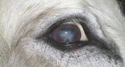
Chronic corneal ulceration – a common finding. Note pigmentation along ventral edge of cornea. (Image courtesy of The Donkey Sanctuary)
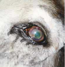
Bandage lens in place. The green spot makes the lens visible. (Image courtesy of The Donkey Sanctuary)
Introduction

The majority of The Donkey Sanctuary’s] donkey population is aged, so corneal integrity is poor.
Clinical Signs
Corneal ulceration is found even with no presenting sign.
Diagnosis
Discovering the cause and resolving the problem is frustratingly difficult. Some general rules are:
- Avoid corticosteroids, systemic or topical
- Auriculopalpebral nerve block is often essential for thorough examination and sampling
- Swab every ulcer for bacteria, and non-healing ulcers for fungi and yeast from the edge of the lesion
- Check for and correct any underlying cause, e.g. entropion, foreign body, viral infection, dry eye. Check behind the third eyelid.
- Ensure feed and management achieve the optimal health conditions. Hypoalbuminaemia causes delay in healing. Ensure dust-free stabling, with hay fed on the ground and mask protection from bright daylight
- Monitor change, using fluorescein dye and diagrams. A deep ulcer involving the stroma will demonstrate less fluorescein uptake. Underrunning of the ulcer edges will be evident using the stain.
Treatment

Deep ulcers can easily rupture and should be treated as an emergency. Sedation, careful handling and local nerve block should be used.
- Antibiotics are used in every case and should be selected on the basis of culture results. Gram-positive bacteria constitute the normal flora but will be the first to invade the damaged cornea. Gram-negative bacteria and fungi are primary invaders and can do severe damage if not targeted specifically
- Topical treatment should be in solution form, as ointments can irritate all but a superficial ulcer
- Anterior uveitis is commonly associated with corneal ulceration. Use atropine drops hourly until miosis resolves and then twice daily for two or three days
- NSAIDs are necessary to relieve initial pain. Flunixin meglumine is most effective in reducing ocular pain, but should be stopped or changed to phenylbutazone as soon as possible due to its adverse effect on cellular repair
- Corneal debridement of all loose epithelium and debris allows repair of the cornea and stimulates the healing response. Auriculopalpebral nerve block and topical anaesthesia facilitate this procedure
- Deep ulcers require surgical repair. Conjunctival grafts are best referred to specialists, who have the appropriate ophthalmic instruments and experience
- Veterinary bandage lenses are useful in reducing discomfort and providing an optimal environment for healing
- Anti-collagenases are still popular despite the lack of proven efficacy. EDTA tubes can be made up with serum, plasma or replacement tears and instilled regularly during healing
- Nasolacrimal or subpalpebral lavage systems are ideal for long-term topical treatments
- Systemic antibiotics are indicated for severe corneal ulcers and stromal infection, but will only deliver to the ulcer if corneal blood vessels have reached the site
References
- Grove, V. (2008) Conditions of the eye In Svendsen, E.D., Duncan, J. and Hadrill, D. (2008) The Professional Handbook of the Donkey, 4th edition, Whittet Books, Chapter 11
|
|
This section was sponsored and content provided by THE DONKEY SANCTUARY |
|---|