Difference between revisions of "Dermatophytosis"
Jump to navigation
Jump to search
m |
m |
||
| Line 8: | Line 8: | ||
}} | }} | ||
<br> | <br> | ||
| − | |||
| − | |||
| − | |||
| − | |||
| − | |||
| − | |||
| − | |||
| − | |||
| − | |||
| − | |||
==General== | ==General== | ||
| Line 139: | Line 129: | ||
*[[Antifungal Drugs]] | *[[Antifungal Drugs]] | ||
| + | |||
| + | {{| align="center" | ||
| + | |<gallery>Image:Dermatophyte arthrospore hair root.jpg|<center><p>Dermatophyte arthrospores on a hair root</p><sup>Copyright Professor Andrew N. Rycroft, BSc, PHD, C. Biol.F.I.Biol., FRCPath</sup></center> | ||
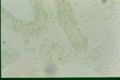
| + | Image:Dermatophyte mycelium in skin.jpg|<center><p>Dermatophytosis mycelium in skin</p><sup>Copyright Professor Andrew N. Rycroft, BSc, PHD, C. Biol.F.I.Biol., FRCPath</sup></center> | ||
| + | Image:Dermatophyte skin KOH.jpg|<center><p>Dermatophyte in skin using a KOH mount</p><sup>Copyright Professor Andrew N. Rycroft, BSc, PHD, C. Biol.F.I.Biol., FRCPath</sup></center> | ||
| + | Image:Dermatophytosis Wood's Lamp.jpg|<center><p>Dermatophytosis lesion in a cat diagnosed using a Wood's Lamp|</p><sup>Copyright Professor Andrew N. Rycroft, BSc, PHD, C. Biol.F.I.Biol., FRCPath</sup></center> | ||
| + | Image:Ringworm on human arm.jpg|<center><p>Ringworm on a human arm</p><sup>Wikimedia Commons</sup></center> | ||
| + | Image:Dermatophytosis in a dog.jpg|<center><p>Dermatophytosis in a dog</p><sup>Copyright Professor Andrew N. Rycroft, BSc, PHD, C. Biol.F.I.Biol., FRCPath</sup></center> | ||
| + | Image:Dermatophytosis in a cow.jpg|<center><p>Dermatophytosis in a cow</p><sup>Copyright Professor Andrew N. Rycroft, BSc, PHD, C. Biol.F.I.Biol., FRCPath</sup></center> | ||
| + | Image:Dermatophytosis in a chicken.jpg|<center><p>Dermatophytosis in a chicken</p><sup>Copyright Professor Andrew N. Rycroft, BSc, PHD, C. Biol.F.I.Biol., FRCPath</sup></center> | ||
| + | Image:Dermatophytosis dog foot.jpg|<center><p>Dermatophytosis lesion on a dog's leg</p><sup>Copyright Professor Andrew N. Rycroft, BSc, PHD, C. Biol.F.I.Biol., FRCPath</sup></center> | ||
| + | Image:Dermatophytosis in a horse.jpg|<center><p>Dermatophytosis in a horse</p><sup>Copyright Professor Andrew N. Rycroft, BSc, PHD, C. Biol.F.I.Biol., FRCPath</sup></center></gallery> | ||
| + | |} | ||
Revision as of 17:27, 4 June 2009
| This article is still under construction. |
|
|
General
- Pigmented, saprophytic organisms called Phaeohyphomycetes
- Previously called 'Fungi Imperfecti'
- The two main species of veterinary interest are Microsporum and Trichophton
- Worldwide
- They are usually secondary invaders
- Able to penetrate all layers of skin, but are generally restricted to the keratin layer and its appendages
- Therefore, most often seen in subcuticular or cutaneous sites
- Lack of tolerance to body temperature and antifungal activity in serum and body fluids prevent the fungi invading subcutaneously
- Transmitted by direct or indirect contact
- Immunuocompromised hosts may develop systemic infections
- Microsporum - zoophilic
- Parasites of animals
- Trichophyton - geophilic
- Inhabits soil
- Epidermophyton - anthropophilic
- Parasites of people
- Common in many species, especially cats
- Hot, humid environment predisposes to infection
- More common in young animals
- Produce proteolytic enzymes to penetrate surface lipid
- Fungal hyphae invade keratin -> break into arthrospores
- Phaeohyphomycosis:
- Occurs sporadically in cats, horses, cattle, fish, reptiles, amphibians, birds, and rarely in dogs
- Examples include: Exophiala sp., Phialophora sp., Pseudomicrodochium sp., Bipolaris sp., Moniella sp., Cladosporium sp., Wangiella sp., Curvularia spp., Exserohilum sp., Alternaria sp., Staphylotrichum sp., and Xylohypha sp
- Culture is necessary for definitive diagnosis
Pathogenesis
- Epidermal hyperplasia (hyperkeratosis, parakeratosis, acanthosis) and inflammation
- Superficial perivascular dermatitis -> exocytosis (migration through epidermal layers) -> intracorneal microabscesses
- Exocytosis -> folliculitis -> furunculosis
- Highly variable lesions
- Normal -> eruptive nodular -> pseudomycetoma -> onychomycosis
- Secondary invasion by Staphylococcus aureus and Staphylococcus intermedius are common and cause pustules in the hair follicles
- Grossly:
- Circular or irregular lesion, may coalesce
- Scaly to crusty patches
- Alopecia due to broken hair shafts and hairs lost from inflammed follicles
- Follicular papules and pustules
- Peripheral red ring (ringworm) due to dead fungi in areas of inflammation at centre of lesions and viable fungi peripherally
- More common in housed animals, rather than animals turned out to pasture
- Highest incidence of disease during the winter
- May resolve spontaneously in the spring and summer
Histology
- Perifolliculitis, folliculitis or furunculosis
- Epidermal hyperplasia
- Intracorneal microabscesses
- Septate hyphae or spores may be found in stratum corneum and keratin of hair follicles
Diagnosis
- Wood's Lamp
- UV light
- Florourescence if fungi present
- Samples can be examined in 10-20% KOH for the presence of hyphae or arthrospores
- Lactophenol Cotton Blue enhances visualisation
- Sabouraud's Dextrose agar containing cyclohexamide and chloramphenicol at room temperature for a month for culture
- Dermatophyte Test Medium
- Saubouraud's Dextrose agar with phenol red indicator
- Medium changes from yellow to red if fungi present
Treatment
- Isolation of infected animal
- Precautions should be taken to prevent human infection
- Griseofulvin best method of treatment
- Expensive
- Oral dosage
- Prolonged treatment required
- Whitfield's ointment
- Salicylic and benzoic acid
- Other treatments:
- Aqueous lime sulphur topically for dogs
- Iodine
- Antibiotics
- Natamycin antifungal
- Imidiazole derivatives
Further Links
- Pathology of dermatophytosis
{{| align="center"
|
Dermatophyte arthrospores on a hair root
Copyright Professor Andrew N. Rycroft, BSc, PHD, C. Biol.F.I.Biol., FRCPath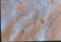
Dermatophytosis mycelium in skin
Copyright Professor Andrew N. Rycroft, BSc, PHD, C. Biol.F.I.Biol., FRCPath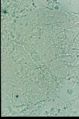
Dermatophyte in skin using a KOH mount
Copyright Professor Andrew N. Rycroft, BSc, PHD, C. Biol.F.I.Biol., FRCPath Copyright Professor Andrew N. Rycroft, BSc, PHD, C. Biol.F.I.Biol., FRCPath
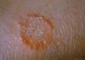
Ringworm on a human arm
Wikimedia Commons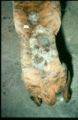
Dermatophytosis in a dog
Copyright Professor Andrew N. Rycroft, BSc, PHD, C. Biol.F.I.Biol., FRCPath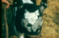
Dermatophytosis in a cow
Copyright Professor Andrew N. Rycroft, BSc, PHD, C. Biol.F.I.Biol., FRCPath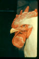
Dermatophytosis in a chicken
Copyright Professor Andrew N. Rycroft, BSc, PHD, C. Biol.F.I.Biol., FRCPath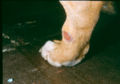
Dermatophytosis lesion on a dog's leg
Copyright Professor Andrew N. Rycroft, BSc, PHD, C. Biol.F.I.Biol., FRCPath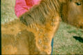
Dermatophytosis in a horse
Copyright Professor Andrew N. Rycroft, BSc, PHD, C. Biol.F.I.Biol., FRCPath


|}