Difference between revisions of "Enzootic Pneumonia - Calves"
m (→Pathology) |
|||
| (41 intermediate revisions by 2 users not shown) | |||
| Line 1: | Line 1: | ||
| − | + | ==Introduction== | |
| + | A pneumonic disease which is caused by a range of infectious agents in combination with management and environmental stress causing damage to the respiratory tract predisposing to disease. | ||
| + | It is principally of viral origin and the following viruses have been implemented in disease. [[Bovine Respiratory Syncytial Virus|Bovine respiratory syncytial virus (BRSV)]], [[Bovine Parainfluenza - 3|Parainfluenza- 3 (PI3)]], [[Bovine Viral Diarrhoea Virus|Bovine viral diarrhoea virus (BVDV)]], [[Bovine Adenovirus|Adenoviruses]], [[Bovine Coronavirus|Calf coronavirus]] and [[Infectious Bovine Rhinotracheitis|Bovine herpes viruses]]. | ||
| + | [[Mycoplasmas]] have also been implemented and secondary bacterial infection is common. | ||
| + | Bacterial agents include [[Mannheimia haemolytica|''Manheimia (Pasteurella) haemolytica'' serotype A1]], [[Pasteurella multocida|''Pasteurella multocida'']], [[Arcanobacter pyogenes|''Arcanobacter pyogenes'']] and ''[[Haemophilus somnus]]''. | ||
| + | All agents are transmitted by aerosol and direct contact. | ||
| − | == | + | ==Signalment== |
| − | + | Mainly a problem in calves less than 6 months old and particularly affects 2-10 week old animals. It occurs more commonly in dairy calves than beef calves. | |
| + | |||
| + | ==Diagnosis== | ||
| + | |||
| + | To establish the cause nasopharangeal swabs or broncho-alveolar lavage can be undertaken and examined for bacteria, viruses or mycoplasma. | ||
| + | |||
| + | Fluorescent antibody tests can be used to detect viral causes. Paired serum samples will also show recent exposure to a viral pathogen. Additionally Post mortem exam can confirm the presence of certain pathogens. | ||
| + | |||
| + | ==History and Clinical signs== | ||
| + | <u>Acutely affected animals:</u> | ||
| + | |||
| + | Affects a group of calves who display respiratory signs including a dry cough, increased respiratory rate, nasal and lacrimal discharge. Animals are pyrexic, depressed and also often anorexic. | ||
| + | |||
| + | <u>Chronically affected animals:</u> | ||
| + | |||
| + | Usually affects a group of animals who are kept indoors. The disease is gradual in onset and although respiratory signs similar to acutely affected animals are present the animals remain bright and eating. | ||
| + | ==Pathology== | ||
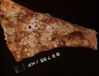
| + | [[Image:Acute suppurative pneumonia.jpg|right|thumb|200px|<small><center>Acute suppurative pneumonia (Image sourced from Bristol Biomed Image Archive with permission)</center></small>]] | ||
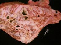
| + | [[Image:Calf pneumonia.jpg|right|thumb|200px|<small><center>Calf pneumonia - chronic, with abscesses, fibrosis (Image sourced from Bristol Biomed Image Archive with permission)</center></small>]] | ||
| + | '''Gross pathology''': | ||
| − | + | Consolidation of the cranioventral lung areas will be present which increases in volume with duration of disease. Exudate is present in the main airway of affected lung lobules with thickening of the surrounding connective tissue. | |
| − | |||
| − | == | + | '''Micro pathology''': |
| − | + | ||
| + | Even though proper follicle formation is present, some of which may be large enough to compress the lumen. | ||
| + | A mixed cell exudate will be present in the airway lumen and substantial lymphoid tissue will be present around the airways. The alveolar walls may be thickened with [[Lymphocytes - Introduction|lymphocytes]]. | ||
| + | |||
| + | ==Treatment and Control== | ||
| + | Once a diagnosis has been made as to the likely causative organisms a number of management issues on the farm must be addressed. These include ensuring each calf ingests enough good quality colostrum, good nutrition, stress management, good housing, and other management of any concurrent diseases. Additionally group sizes should be assessed and ideally no more than 20 calves should be housed per group. Animals should be kept in groups of the same age and should not share airspace with adult cattle. If this is not possible animals should be arranged with the air flowing from youngest to oldest. Isolation/ hospital pens should be available to prevent spread of disease and to ensure affected animals are cared for correctly. | ||
| + | Additionally, once causative organisms have been identified [[Vaccines|vaccination]] programmes can also be put in place for cows 4 weeks pre-partum to improve colostral antibodies that the calves will receive. | ||
| + | |||
| + | ==Prognosis== | ||
| + | Good if recognised early and if affected animals are treated and management is improved. | ||
| + | |||
| + | ==Literature Search== | ||
| + | [[File:CABI logo.jpg|left|90px]] | ||
| + | |||
| + | |||
| + | Use these links to find recent scientific publications via CAB Abstracts (log in required unless accessing from a subscribing organisation). | ||
| + | <br><br><br> | ||
| + | [http://www.cabdirect.org/search.html?rowId=1&options1=AND&q1=%22Enzootic+Pneumonia%22&occuring1=title&rowId=2&options2=AND&q2=cattle&occuring2=od&rowId=3&options3=AND&q3=&occuring3=freetext&x=40&y=17&publishedstart=yyyy&publishedend=yyyy&calendarInput=yyyy-mm-dd&la=any&it=any&show=all Enzootic pneumonia of calves publications] | ||
| + | |||
| + | ==References== | ||
| + | Andrews, A.H, Blowey, R.W, Boyd, H and Eddy, R.G. (2004) '''Bovine Medicine''' (Second edition), ''Blackwell Publishing'' | ||
| + | |||
| + | Merck & Co (2008) '''The Merck Veterinary Manual''' (Eighth Edition) ''Merial '' | ||
| − | |||
| − | + | {{review}} | |
| − | + | [[Category:Respiratory Diseases - Cattle]] | |
| − | |||
| − | |||
| − | |||
| − | |||
| − | |||
| − | |||
| − | |||
| − | |||
| − | |||
| − | |||
| − | |||
| − | |||
| − | |||
| − | |||
| − | |||
| − | |||
| − | |||
| − | |||
| − | |||
| − | |||
| − | |||
| − | |||
| − | |||
| − | |||
| − | |||
| − | |||
[[Category:Respiratory_Bacterial_Infections]] | [[Category:Respiratory_Bacterial_Infections]] | ||
| − | [[Category: | + | |
| + | |||
| + | [[Category:Brian Aldridge reviewing]] | ||
Latest revision as of 20:29, 5 March 2014
Introduction
A pneumonic disease which is caused by a range of infectious agents in combination with management and environmental stress causing damage to the respiratory tract predisposing to disease. It is principally of viral origin and the following viruses have been implemented in disease. Bovine respiratory syncytial virus (BRSV), Parainfluenza- 3 (PI3), Bovine viral diarrhoea virus (BVDV), Adenoviruses, Calf coronavirus and Bovine herpes viruses. Mycoplasmas have also been implemented and secondary bacterial infection is common. Bacterial agents include Manheimia (Pasteurella) haemolytica serotype A1, Pasteurella multocida, Arcanobacter pyogenes and Haemophilus somnus. All agents are transmitted by aerosol and direct contact.
Signalment
Mainly a problem in calves less than 6 months old and particularly affects 2-10 week old animals. It occurs more commonly in dairy calves than beef calves.
Diagnosis
To establish the cause nasopharangeal swabs or broncho-alveolar lavage can be undertaken and examined for bacteria, viruses or mycoplasma.
Fluorescent antibody tests can be used to detect viral causes. Paired serum samples will also show recent exposure to a viral pathogen. Additionally Post mortem exam can confirm the presence of certain pathogens.
History and Clinical signs
Acutely affected animals:
Affects a group of calves who display respiratory signs including a dry cough, increased respiratory rate, nasal and lacrimal discharge. Animals are pyrexic, depressed and also often anorexic.
Chronically affected animals:
Usually affects a group of animals who are kept indoors. The disease is gradual in onset and although respiratory signs similar to acutely affected animals are present the animals remain bright and eating.
Pathology
Gross pathology:
Consolidation of the cranioventral lung areas will be present which increases in volume with duration of disease. Exudate is present in the main airway of affected lung lobules with thickening of the surrounding connective tissue.
Micro pathology:
Even though proper follicle formation is present, some of which may be large enough to compress the lumen. A mixed cell exudate will be present in the airway lumen and substantial lymphoid tissue will be present around the airways. The alveolar walls may be thickened with lymphocytes.
Treatment and Control
Once a diagnosis has been made as to the likely causative organisms a number of management issues on the farm must be addressed. These include ensuring each calf ingests enough good quality colostrum, good nutrition, stress management, good housing, and other management of any concurrent diseases. Additionally group sizes should be assessed and ideally no more than 20 calves should be housed per group. Animals should be kept in groups of the same age and should not share airspace with adult cattle. If this is not possible animals should be arranged with the air flowing from youngest to oldest. Isolation/ hospital pens should be available to prevent spread of disease and to ensure affected animals are cared for correctly. Additionally, once causative organisms have been identified vaccination programmes can also be put in place for cows 4 weeks pre-partum to improve colostral antibodies that the calves will receive.
Prognosis
Good if recognised early and if affected animals are treated and management is improved.
Literature Search
Use these links to find recent scientific publications via CAB Abstracts (log in required unless accessing from a subscribing organisation).
Enzootic pneumonia of calves publications
References
Andrews, A.H, Blowey, R.W, Boyd, H and Eddy, R.G. (2004) Bovine Medicine (Second edition), Blackwell Publishing
Merck & Co (2008) The Merck Veterinary Manual (Eighth Edition) Merial
| This article has been peer reviewed but is awaiting expert review. If you would like to help with this, please see more information about expert reviewing. |


