|
|
| Line 5: |
Line 5: |
| | | | |
| | ==[[Equine Nose - Horse Anatomy|Nose]]== | | ==[[Equine Nose - Horse Anatomy|Nose]]== |
| − | Olfaction is the sense of smell, which is the ability to perceive and distinguish odours. The sense of smell is well developed in horses, as they are prey animals.
| |
| − | [[Image:Nasal Cavities.jpg|thumb|right|250px|Nasal Cavities - Copyright David Bainbridge]]
| |
| − | The nose consists of the external nares with nasal cartilages, the [[Equine Upper Respiratory Tract - Horse Anatomy#Nasal Cavity|nasal cavity]] (including the nasal meatus and conchae), and the [[Equine Upper Respiratory Tract - Horse Anatomy#Paranasal Sinuses|paranasal sinuses]]. The borders of the nasal cavity are as follows:
| |
| − |
| |
| − | '''Caudal''': The cribrifrom plate of the ethmoid bone.
| |
| − |
| |
| − | '''Ventral''': Continuous with the nasopharynx.
| |
| − |
| |
| − | '''Dorsal''': The maxilla and the palatine processes of the incisive bones.
| |
| − |
| |
| − | Rostrally, the median septum is a continuation of the ethmoid bone. The median septum is made up of hyaline cartilage, and divides the [[Equine Upper Respiratory Tract - Horse Anatomy#Nasal Cavity|nasal cavity]] into left and right halves.
| |
| − |
| |
| − | ===Nasal conchae===
| |
| − |
| |
| − | Nasal conchae are '''turbinate bones''' that project into the nasal cavity. Their purpose is to increase the respiratory surface area, and to create turbulence within the passing air. This helps to filtrate and warm or cool the air that passes through. They are cartilagenous or ossified scrolls that are covered with mucous membrane, under which is a layer of anastomosing blood vessels. There are dorsal and ventral conchae, the dorsal concha extending further into the nasal cavity. The conchae divide the nasal cavity into meatuses, which branch out from a common nasal meatus which is adjacent to the nasal septum. There are three nasal meatuses which branch from the common nasal meatus: '''dorsal''', '''middle''' and '''ventral''':
| |
| − |
| |
| − | '''Dorsal nasal meatus''': The passage between the roof of the nasal cavity and the dorsal nasal concha.
| |
| − |
| |
| − | '''Middle nasal meatus''': Between the dorsal and ventral conchae, and communicates with the [[Equine Upper Respiratory Tract - Horse Anatomy#Paranasal Sinuses|paranasal sinuses]].
| |
| − |
| |
| − | '''Ventral nasal meatus''': The main pathway for airflow leading to the [[Pharynx - Anatomy & Physiology|pharynx]], and is positioned between the ventral nasal concha and the floor of the [[Equine Upper Respiratory Tract - Horse Anatomy#Nasal Cavity|nasal cavity]].
| |
| − |
| |
| − | '''Common nasal meatus''': The longitudinal space on either side of the nasal septum.
| |
| − |
| |
| − | The [[Equine Upper Respiratory Tract - Horse Anatomy#Paranasal Sinuses|paranasal sinuses]] are extensions of the [[Equine Upper Respiratory Tract - Horse Anatomy#Nasal Cavity|nasal cavity]].
| |
| − |
| |
| − | ===Vasculature===
| |
| − | There is a dense network of blood vessels supplying the nasal mucosa. The '''sphenopalatine artery''' supplies the [[Equine Upper Respiratory Tract - Horse Anatomy#Nasal Cavity|nasal cavity]]. The lateral and dorsal '''nasal arteries''', which are branches of the facial artery, itself a branch of the '''external carotid artery''', supply the [[Equine Upper Respiratory Tract - Horse Anatomy#Nose|nose]]. The '''infraorbital artery''' also supplies the [[Equine Upper Respiratory Tract - Horse Anatomy#Nose|nose]].
| |
| − |
| |
| − | ===Innervation===
| |
| − | Sensory innervation is provided by the '''olfactory nerve''' ([[Equine Cranial Nerves - Horse Anatomy#Olfactory Nerve (I)|CN I]]).
| |
| − |
| |
| − | ===Olfaction===
| |
| − | [[Image:Olfactory Epithelium.jpg|thumb|right|250px|Olfactory Epithelium - Copyright David Bainbridge]]
| |
| − | The '''olfactory sensory neurones''' are present in the olfactory epithelium. It is the mucous membrane of the dorsal nasal conchae that is sensitive to smell. Here, odourants dissolve in the mucous membrane, and it is these odourants that are recognised by the olfactory sensory receptors. '''Sensory cilia''', that are present on the surface of the olfactory receptors, project into the film of mucous. The mucous also contains antibodies, to prevent infection. This is because the olfactory neurones provide a direct passage, via the olfactory nerve ([[Equine Cranial Nerves - Horse Anatomy#Olfactory Nerve (I)|CN I]]), to the [[Equine Brain - Horse Anatomy|brain]].
| |
| − |
| |
| − | ===Central Olfactory Pathways===
| |
| − | [[Image:Central Olfactory Pathways.jpg|thumb|right|250px|Central Olfactory Pathways - Copyright David Bainbridge]]
| |
| − | The olfactory receptors are embedded in the mucous membrane within the [[Equine Upper Respiratory Tract - Horse Anatomy#Nasal Cavity|nasal cavity]]. They are '''bipolar neurones''', and are covered with cilia (non-motile). It is thought that these cilia contain the active sites for the olfactory transduction process. The axons from the olfactory receptors join together and become the olfactory nerve ([[Equine Cranial Nerves - Horse Anatomy#Olfactory Nerve (I)|CN I]]). The axons pass through the perforations in the '''cribriform plate''' of the ethmoid bone, and enter the '''olfactory bulb'''. Once the axons have entered the olfactory bulb, the olfactory nerve synapses on '''mitral cells'''. The axons from these cells then project into the '''olfactory cortex''' of the [[Equine Brain - Horse Anatomy#Forebrain|cerebral hemispheres]], via the '''olfactory tract'''. The olfactory cortex is the only region within the [[Equine Brain - Horse Anatomy#Forebrain|cerebral hemispheres]] that receives direct sensory input without any information first passing through the [[Equine Brain - Horse Anatomy#Forebrain|thalamus]]. This is because the olfactory system evolved before the [[Equine Brain - Horse Anatomy#Forebrain|thalamus]].
| |
| − |
| |
| − | ===Vomeronasal Organ===
| |
| − |
| |
| − | Click here for information on the [[Vomeronasal Organ]].
| |
| − |
| |
| − | ===Histology===
| |
| − | Olfactory epithelium is made up of '''olfactory cells''', '''sustentacular cells''' and '''basal cells'''. The olfactory axons and olfactory glands are present in the lamina propria. The first part of the [[Equine Upper Respiratory Tract - Horse Anatomy#Nasal Cavity|nasal cavity]], from the nostrils, is lined by keratinised stratified squamous epithelium. Sebaceous glands and hairs are also present in this region. The hairs function to keep dust out of the [[Equine Upper Respiratory Tract - Horse Anatomy#Nasal Cavity|nasal cavity]]. The upper part of the [[Equine Upper Respiratory Tract - Horse Anatomy#Nasal Cavity|nasal cavity]]is also lined by stratified squamous epithelium, but it is no longer keratinised. The epithelium then becomes pseudostratified columnar, and is ciliated. This is typical of the respiratory system, so this type of epithelium may also be known as respiratory epithelium. Goblet cells, which produce mucous, are present in this region of the nasal cavity. Mucous and serous glands are present in the connective tissue, the lamina propria, which lies underneath the epithelium that covers the nasal conchae. The olfactory region of the nasal cavity is formed by the membrane covering the dorsal nasal conchae. Cilia in this region are formed from olfactory cells, although they are non-motile. It is the membrane that covers these cilia that contains the olfactory receptors that are sensitive to smell, after the odours have been dissolved in the serous membrane covering the epithelium. The axons of these receptors bundle together within the lamina propria. The olfactory cells gain mechanical and metabolic support from the sustentacular cells.
| |
| − |
| |
| − | ===Links===
| |
| − | *Click here for more information on the [[Equine Upper Respiratory Tract - Horse Anatomy#Nasal Cavity|nasal cavity]]
| |
| − | *Click here for more information on the [[Equine Upper Respiratory Tract - Horse Anatomy#Paranasal Sinuses| paranasal sinuses]]
| |
| − | *Click here for more information on [[Diseases of the nasal cavity and sinuses|diseases of the nasal cavity and sinuses]].
| |
| | | | |
| | ==Gustatory System== | | ==Gustatory System== |
Template:Incomplete
Gustatory System
Gustation is the sense of taste, and is a system involving chemoreceptors. The gustatory system can usually detect four different types of taste: bitter, sweet, sour and salt. These tastes are detected by taste buds that are contained within papillae, which are mainly found on the dorsal surface of the tongue. These tastes are relayed from the taste buds, via the olfactory nerve (CN I), to the brain.
The tongue is the main structure involved in taste. The tongue is covered by a lingual mucosa, which is tough, and most of its surface is covered with papillae. The papillae are a local modifictaion of the lingual mucosa. There are also a few taste buds present on the epiglottis and the pharynx. They are grouped according to their function: mechanical papillae are cornified and protect the deeper structures of the tongue, and gustatory papillae, which are covered in taste buds.
| Group Name
|
Types of Papillae
|
Function
|
| Mechanical Papillae
|
1. Filiform papillae
2. Conical papillae
3. Marginal papillae
|
1. The smallest and most numerous papillae
2. Larger, but less numerous; plentiful over the dorsal
surface of the tongue of the ox and cat, this
being the reason that their tongues are rough
3. Present in new-born carnivores and piglets,
and help with suckling
|
| Gustatory Papillae
|
1. Fungiform papillae
2. Vallate papillae
3. Foliate papillae
|
Taste buds are contained within the epithelium
of the gustatory papillae, the taste buds being
sensitive to taste
|
See the link for further information on types of papillae: The Tongue and Taste Buds
There are salivary glands in the regions of the taste buds. These salivary glands remove small particles of food from the papillae, to make the papillae free for new food entering the mouth.
Taste Buds
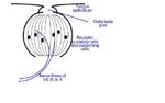
Taste Bud - Copyright David Bainbridge
Taste buds are made up of a group of eptihelial cells, and are contained within papillae. They contain three major cell types:
1. Supporting (sustentacular) cells - these cells mainly form the outer layer of the taste bud
2. Gustatory cells - these cells are chemoreceptors, and are located in the centre of the taste bud
3. Basal cells
The soft palate, pharynx and nasal cavity contribute to taste sensation, mainly due to olfactory information. Other factors that contribute to taste are consistency and temperature of food. Taste buds can detect four different types of taste: salt, sweet, bitter and sour. There are no structural differences among the taste buds that detect these different types of taste. A taste receptor is a chemoreceptor that allows taste. There are two types of taste receptor:
1. Salt and sour (acid): ion channels
2. Bitter and sweet: G-protein coupled receptors (GPCRs) and ion channels
Each taste receptor allows a different sort of sensory transduction. After the taste receptors have detected the presence of a certain compound, they start an action potential which reaches the brain. These action potentials are conveyed to the brain via three of the cranial nerves:
- Facial nerve (CN VII): carries action potentials from the rostral two-thirds of the tongue
- Glossopharyngeal nerve (CN XII): carries action potentials from the caudal third of the tongue
- Vagus nerve (CN X): carries some of the action potentials from the back of the oral cavity
Sensory neurones synapse in the solitary nucleus of the medulla.
Vasculature
The main blood supply to the tongue is via the lingual artery, which is a branch of the external carotid artery. A secondary blood supply to the tongue is provided via the tonsillar branch of the facial artery and the ascending pharyngeal artery.
Innervation
The rostral 2/3 of tongue is innervated by the lingual branch of the trigeminal nerve (CN V), which is sensory supplying temperature, touch and pain. The chorda tympani of the facial nerve (CN VII) supplies the taste. The caudal 1/3 of tongue is innervated by the glossopharyngeal (CN IX), providing motor function for taste.
Central Gustatory Pathways
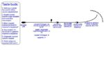
Central Gustatory Pathways - Copyright David Bainbridge
Receptor cells have a single receptor type, yet afferent nerve fibres carry information from several different cell types. This means that the brain has to re-discriminate between the tastes by cross-comparison between inputs from many fibres.
Salt
There is an ion channel in the wall of the taste bud cells, which allows sodium ions (Na+) to enter the cell. This causes depolarisation of the cell, which causes the opening of voltage regulated calcium ion (Ca2+) gates, causing calcium enters to flood into the cell, which then causes the release of a neurotransmitter.
Sweet
Sweet tastes are conveyed via G-protein coupled receptors (GPCRs). Sweet compounds such as saccharides activate the GPCR, which causes the release of a substance called gustducin, which itself then activates a molecule called adenylate cyclase, which is present inside the cell. Adenylate cyclase causes an increase in the concentration of the molecule cAMP, which itself will cause the closure of potassium ion (K+) channels. This will lead to depolarisation, and then the release of a neurotransmitter.
Bitter
Bitter tastes are conveyed via G-protein coupled receptors (GPCRs). Bitter compounds activate the GPCR, which causes the release of a substance called gustducin. Gustducin is made up of three subunits, which, when activated by the GPCR, break apart and activate a local enzyme, phosphodiesterase. Phosphodiesterase then converts a precursor within the cell into a secondary messenger, which itself causes the closure of potassium ion (K+) channels. The secondary messenger can also stimulate the endoplasmic reticulum to release calcium ions (Ca2+), which help to cause depolaristion. Depolarisation leads to accummulation of potassium ions within the cell, then depolarisation, which leads to release of a neurotransmitter.
Sour
Sour taste indicates the presence of acidic compounds. Three different recptors are present for the detection of sour taste:
1. An ion channel that allows hydrogen (H+) ions to flow into the cell.
2. A potassium ion (K+) channel, which allows potassium ions to escape from the cell. These channels are blocked by hydrogen ions, so the potassium ions are trapped inside the cell.
3. A protein which opens to sodium (Na+) ions when a hydrogen (H+) ion attaches to it. This allows sodium ions to flow down its concentration gradient into the cell. This influx allows opening of the voltage regulated calcium ion (Ca2+) gates
These receptors work together, leading to depolarisation of the cell, which then leads to the release of a neurotransmitter.
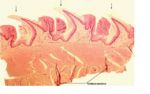
Histology
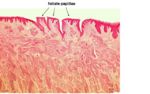
Filiform Papillae - Copyright John Bredl
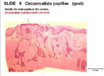
Circumvallate Papillae - Copyright John Bredl
Foliate Papillae - Copyright John Bredl
The tongue is lined by stratified squamous epithelium.
- Filiform Papillae: no glands, no taste buds, no lymphatic tissue.
- Circumvallate Papillae: contain glands, taste buds and lymphatic tissue. The glands open into the moat around the papillae. The taste buds are present on the sides of the papillae. The lymphatic tissue is found deeper into the papillae.
- Foliate Papillae: form a series of parallel folds. They contain glands, taste buds and some lymphatic tissue. The glands are found deep inside and between the papillae. The taste buds are found on the sides of the papillae.


