Difference between revisions of "Female Reproductive Tract - Horse Anatomy"
(→Uterus) |
(→Uterus) |
||
| Line 113: | Line 113: | ||
====Anoestrus==== | ====Anoestrus==== | ||
The endothelium is relatively thin with little endometrial proliferation or gland development. This is due to the absence of the ovarian steroids oestradiol and progesterone. The glands are lined by '''simple cuboidal epithelium'''. | The endothelium is relatively thin with little endometrial proliferation or gland development. This is due to the absence of the ovarian steroids oestradiol and progesterone. The glands are lined by '''simple cuboidal epithelium'''. | ||
| + | ===Innervation=== | ||
| + | The uterus is innervated by both sympathetic and parasympathetic fibres, which play a part in the regulation of uterine activity. | ||
| + | ==Vasculature== | ||
| + | *'''Uterine branch of the ovarian artery''' supplies the cranial parts of the uterine horns. | ||
| + | *'''Uterine artery''' supplies the rest of the uterine horns and the uterine body. This is a branch off the '''External Iliac artery''' in the mare. The '''Uterine artery''' and the '''Ovarian artery''' anastomose within the [[Broad Ligament - Anatomy & Physiology|Broad Ligament]]. | ||
==Cervix== | ==Cervix== | ||
==Vagina and Vestibule== | ==Vagina and Vestibule== | ||
Revision as of 15:59, 26 November 2012
| This article is still under construction. |
Ovaries
The ovary is the female Gonad homologous to the male Testes.The structures found within the ovary are undergoing constant changes throughout the oestrus cycle from the Follicles containing Oocytes, to the formation of Corpus Haemorrhagicum, Corpus Luteum, and finally Corpus Albicans. The ovaries of the mare are two kidney-shaped structures situated ventral to the 4th and 5th lumbar vertebrae, supported by the mesovarium. During the non-breeding season they measure 2-4cm in length and 2-3cm in width. During the sexually active stage, they are 6-8cm in length and 3-4cm in width.

In the mare, the structure of the ovary is reversed compared to other species. The cortex is in the centre and is enclosed by a dense, richly vascularised connective tissue layer, which is analagous to the medulla of other domestic mammals.
Tunica Albiguinea
The connective tissue layer covering the ovarian cortex. The whole ovary is contained within the tunica albiguinea except the ovulation fossa. Overlying this structure is a single layered germinal epithelium.
Cortex
Contains Follicles in various stages of development as well as the Corpus Luteum (includes Corpus Haemorrhagicum and Corpus Albicans). The cortex reaches the surface of the ovary at the ovulation fossa, a deep indentation at the free margin. This is where mature follicles rupture in ovulation, as opposed to at various points on the surface in other domestic mammals. Follicles can be identified by transrectal palpation, but Corpora Lutea cannot. Identification of Corpora Lutea requires ultrasonography.
Medulla
The Medulla is made up of dense connective tissue. This is where all of the lymphatics, nerves and vasculature of the ovary are found.
Histology
Stroma
The body of the ovary (ovarian stroma) consists of:
- Spindle-shaped cells
- Fine collagen fibres
- Ground substance
Stromal cells resemble fibroblasts, but some contain lipid droplets. Bundles of smooth muscle cells are scattered throughout the stroma.
Cortex
Contains:
- Follicles containing oocytes in various stages of development
- Atretic Follicles
- Corpora lutea
- Corpora albicans
The superficial cortex is more fibrotic than the deep, and is called the tunica albuginea. On the surface of the ovary is the germinal epithelium. This is a continuation of the peritoneum.
Medulla
The medulla is highly vascular. It contains hilus cells, which are similar to the Leydig cells of the testes.
Vasculature
Arterial Supply
- The ovarian artery (a branch of the Aorta) and ovarian branches of the uterine artery form anastomoses in the mesovarium and the broad ligament. From this arterial plexus ~10 coiled helicine arteries enter the hilus of the ovary. Smaller branches form a plexus at the corticomedullary junction, giving rise to straight cortical arterioles, which radiate into the cortex. Here they branch and anastomose to form vascular arcades, which give rise to a rich capillary network around follicles.
Venous Drainage
Venous drainage follows the course of the arterial system. Medullary veins are large and tortuous. The ovarian artery is closely associated with the uterine vein. This is important for the transfer of luteolytic PGF2α from the uterus to the ovary.
Lymphatics
Arise in the perifollicular stroma and drain into larger vessels, which coil around the medullary veins.
Innervation
Sympathetic fibres of the autonomic nervous system supply blood vessels and terminate on smooth muscle cells in the stroma around follicles. They may play a role in follicular maturation and ovulation, but the main control is via the endocrine system.
Function
The ovary has two main functions:
- Producing the female gametes oocytes via Gametogenesis.
- Producing the reproductive hormones Oestrogens and Progesterone, an endocrine function.
Processes Taking Place In The Ovary
Additional Learning Resources
Oviducts
The Oviduct is the tube that links the ovary to the uterus and which the ovulated oocyte travels down to become fertilised by sperm present in the female tract. It is also refered to as the Fallopian tube, Uterine tube or Ovarian tube. The Oviduct is suspended from the abdominal wall by the mesosalpnix of the broad ligament. It functions to provide regulation check points for unfertilised oocytes. The mucosal glands produce oviduct secretions integral for:
- Supporting the unfertilised oocyte
- Supporting spermatozoa in the oviduct
- Increasing the fertilising capabilities of the spermatozoa
- Development of the early embryo
The oviduct is devided into 3 anatomical regions:
Infundibulum
The cranial ovarian end of the oviduct. It comprises of numerous fimbrae and the opening into the oviduct tube, the ostium.
Ampulla
The longest region of the oviduct, occupying more than half of its total length, and also has the largest diameter. This is the site of fertilisation. It is distinguished by its many mucosal folds. The ampulla is joined to the isthmus via the Ampullary-Isthmus junction. This junction is important in the mare, as it acts as a regulatory checkpoint allowing only fertilised ova to pass any further along the oviduct and into the uterus.
Isthmus
The caudal end of the oviduct joined to the uterus. The Isthmus is thicker walled than the ampulla and smaller in diameter. Its folded mucosa forms a functional reservoir for sperm in the female tract. Sperm in the female tract reach the isthmus of the oviduct and bind to the mucosal epithelial cells, forming a functional reservoir. The sperm are only released from the isthmus mucosa by the action of paracrine signals from an ova travelling down the oviduct. The formation of a functional reservoir is a possible supporting mechanism of block to polyspermy, as only a few sperm are released from the isthmus mucosa at any one time. This results in only a few sperm being in the vicinity of the ova at a time and so in a position of fertilising the ova.
Uterotubal Junction
The oviducts open into the uterine horn through the uterine ostium. This marks the site of the uterotubal junction. This junction is gradual in ruminants and pigs, but abrupt in the horse and carnivores. In the horse, the uterine ostium is located on top of a papilla, which forms a barrier against ascending infections.
Histology
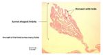
Histological Section of the Oviduct to show Fimbriae of the Infundibulum- Courtesy of J.Bredl, Copyright RVC 2008
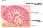
Histological Cross-Section of the Oviduct to show the Ampulla- Courtesy of J.Bredl, Copyright RVC 2008
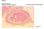
Histological Cross-Section of the Oviduct to show the Isthmus- Courtesy of J.Bredl, Copyright RVC 2008


Infundibulum
Fimbriae are finger-like projections that aid the infundibulum in gliding over the surface of the ovary.
Ampulla
- Ciliated Columna Epithelium
- Thin muscularis layer
- Fern-like mucosal folds
Isthmus
- Simple columnar epithelium
- Thick muscularis layer divided into inner circular layer and outer longditudinal layer
- Few mucosal folds
Vasculature
Tubalar branch of the ovarian artery.
Uterus
The uterus is the organ of pregnancy, as this is where implantation and development of the feotus occurs. The uterus is the reproductive organ with the most species variations. These variations occur in both the anatomical types of uterus as well as the uterine horn appearance and endometrial linings.

The mare has a bicornuate uterus, consisting of a large body and two divergent horns. The uterine body and horns are approximately equal length. The uterus is located within the abdominal cavity, dorsal to the intestines. The uterus is attached to the dorsal body wall via the mesometrium.
The uterus provides a suitable environment for embryo development and attachment. The secretions produced by the endometrial glands are important for maintaining the preimplantion embryo. In response to increasing amounts of oxytocin production by the corpus luteum during the luteal phase, the endometrium produces luteolytic PGF2a to cause degeneration of the corpus luteum if the female is not pregnant. The uterus contributes varying amounts of maternal tissues towards the placenta. The myometrium is involved with sperm transport through the uterus towards the oviduct. Contractions of the myometrium during parturition is important for feotus and placenta expulsion.
Perimetrium
Serosal connective tissue layer continuous with the Broad Ligament.
Myometrium
Muscularis layer made up from thick inner circular smooth muscle layer and thin outer longitudinal muscle layer. The circular and longitudinal layers are seperated by the Stratum Vasculare, a connective tissue layer containing the blood vessels and nerves of the uterine wall.
Endometrium
Mucosa & Submucosa layer containing endometrial glands. The surface of the endometrium has uterine folds in the mare.
Histology
The appearance of the uterus varies with the stages of the oestrus cycle.
Follicular phase-Oestrus
Increasing numbers of uterine glands developing and elongating within the endometrium due to the influence of oestrogens produced by the developing follicles. There is a general increase in the thickness of the endometrium. The epithelium lining the glands is simple columnar epithelium.
Luteal phase
The endometrium is at its maximum thickness with a large number of highly developed glands. This is the secretory phase for the endometrium.
Anoestrus
The endothelium is relatively thin with little endometrial proliferation or gland development. This is due to the absence of the ovarian steroids oestradiol and progesterone. The glands are lined by simple cuboidal epithelium.
Innervation
The uterus is innervated by both sympathetic and parasympathetic fibres, which play a part in the regulation of uterine activity.
Vasculature
- Uterine branch of the ovarian artery supplies the cranial parts of the uterine horns.
- Uterine artery supplies the rest of the uterine horns and the uterine body. This is a branch off the External Iliac artery in the mare. The Uterine artery and the Ovarian artery anastomose within the Broad Ligament.