Difference between revisions of "Fertilisation - Anatomy & Physiology"
Jump to navigation
Jump to search
| Line 9: | Line 9: | ||
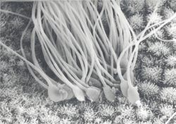
[[Image:Fertilization histology.jpg|right|thumb|250px|<small><center> Electron Scanning Micrograph of Spermatozoa in the Uterine Horn. Copyright RVC 2008 (Courtesy of John Bredl (RVC))</center></small>]] | [[Image:Fertilization histology.jpg|right|thumb|250px|<small><center> Electron Scanning Micrograph of Spermatozoa in the Uterine Horn. Copyright RVC 2008 (Courtesy of John Bredl (RVC))</center></small>]] | ||
| + | |||
| + | |||
| + | |||
| + | |||
| + | |||
| + | |||
| + | |||
| + | |||
| + | |||
Revision as of 14:49, 3 July 2008
Fusion with the Oocyte
- When the Spermatozoan completely penetrates the Zona Pellucida and reaches the Perivitelline Space, it settles into a bed of microvilli formed by the Oocyte plasma membrane.
- Oocyte plasma membrane fuses with the equitorial segment and the fertilizing Spermatozoon is engulfed.
- Brought about by a fusion protein that is inactive prior to the acrosome reaction.
- Nucleus of the Spermatozoon is within the Oocyte cytoplasm.
- Sperm nuclear membrane disappears.
- Sperm nucleus decondenses.
Cortical Reaction - Block to Polyspermy
- During the first and second meiotic divisions of Oogenesis small,dense cortical granules move to the periphery of the Oocyte cytoplasm.
