Difference between revisions of "Hiatal Hernia"
JamesSwann (talk | contribs) |
JamesSwann (talk | contribs) |
||
| Line 16: | Line 16: | ||
<gallery> | <gallery> | ||
| − | Image:Shar_pei.jpg|'''Shar Pei'''<p> | + | Image:Shar_pei.jpg|'''Shar Pei'''<p><small> Copyright Wrinkle 2009 Wikimedia Commons |
| − | Image:Chow_chow.jpg|'''Chow Chow'''<p> | + | Image:Chow_chow.jpg|'''Chow Chow'''<p><small> Copyright Pleple2000 2007 Wikimedia Commons |
| − | Image:Bulldog.jpg|'''Bulldog'''<p> | + | Image:Bulldog.jpg|'''Bulldog'''<p><small> Copyright Makemi 2006 Wikimedia Commons |
| − | Image:French_bulldog.jpg|'''French Bulldog'''<p> | + | Image:French_bulldog.jpg|'''French Bulldog'''<p><small> Copyright danny O 2007 Wikimedia Commons |
</gallery> | </gallery> | ||
Revision as of 11:43, 6 July 2010
| This article is still under construction. |
Description
A hiatal hernia is an abnormality of the diaphragm that allows part of the stomach and the abdominal oesophagus to displace into the thoracic cavity. Two types of hiatal hernia have been recognised in the dog and cat:
- Sliding hiatal hernia - Cranial displacement of the distal oesophagus and stomach into the mediastinum through the oesophageal hiatus of the diaphragm. This is the most common form and it can occur in the dog and cat as a congenital or acquired lesion. Congenital hernias result from the incomplete fusion of the septum transversum (which forms the diaphragm) during early embryonic development.
- Para-oesophageal or Rolling hiatal hernia - Cranial displacement of the gastric fundus into mediastinum through the oesophageal hiatus but adjacent to the oesophagus and gastric cardia which remain in their normal positions. This form of hernia is rare in animals.
Acquired hernias can occur in any breed of dog or cat and these often occur with disorders that cause increases in intra-abdominal pressure (such as chronic vomiting) or increases in negative intrathoracic pressure (such as intermittent airway obstruction seen with laryngeal paralysis or brachycephalic obstructive airway syndrome (BOAS)).
Signalment
Breeds of dog that develop congenital sliding hernias include Chinese Shar-peis and Chow-chows whereas brachycephalic dogs (such as English and French bulldogs) ofetn develop acquired hernias.
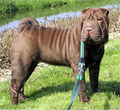 Shar Pei
Copyright Wrinkle 2009 Wikimedia Commons
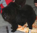 Chow Chow
Copyright Pleple2000 2007 Wikimedia Commons
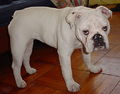 Bulldog
Copyright Makemi 2006 Wikimedia Commons
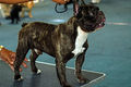 French Bulldog
Copyright danny O 2007 Wikimedia Commons


Diagnosis
Clinical Signs
Some animals may be asymptomatic but otherwise clinical signs include:
- Regurgitation due to impaired function of the herniated lower oesophageal sphincter.
- Hypersalivation related to regurgitation
- Dyspnoea and coughing if the hernia is large and impinges on the lungs or if the animal develops aspiration pneumonia as a result of regurgitation.
- Dehydration and weight loss due to chronic regurgitation.
Diagnostic Imaging
- Plain survey radiographs may show a gas-filled soft tissue opacity in the caudodorsal thorax, continuous with the diaphragmatic margin. Secondary megaoesophagus may develop in longstanding cases and an alveolar lung pattern may be apparent, especially cranio-ventrally, if the animal is developing aspiration pneumonia.
- Barium contrast studies may be used to confirm a diagnosis.
Intermittent hiatal hernias can be difficult to detect and therefore it is sometimes necessary to put pressure on the abdomen during radiography to induce displacement of the stomach.
- Fluoroscopy can be used to identify cases of intermittent herniation if the condition is still suspected after plain radiography.
- Endoscopy may demonstrate cranial displacement of the lower oesophageal sphincter and a large oesophageal hiatus.
Treatment
If the hernia is acquired, the underlying cause should be treated.
Medical management should be initiated to reduce oesophagitis caused by regurgitation. Medical management can be continued for cases with acquired hernias and may also achieve some success in cases with congenital hernias. This approach involves the use of:
- Gastroprotective Drugs including oral sucralfate suspensions and gastric acid secretory inhibitors such as cimetidine, ranitidine or omeprazole.
- A low fat diet fed from a height will increase the tone of the lower oesophageal sphincter and increase the speed of gastric emptying, reducing the likelihood of regurgitation.
- Metaclopramide may also be used to increase the tone of the lower oesophageal sphincter.
- Antibiotics, nebulisation and coupage may be used to manage aspiration pneumonia.
Surgical management is usually attempted with congenital cases (after medical management has been attempted) and to treat the underlying cause in acquired cases.
- Hernia repair is achieved via a cranial ventral coeliotomy. The oesophageal hiatus is exposed by transection of left triangular ligament (between the liver and diaphragm) and retraction of the liver. The phreno-oesophageal ligament is partially incised and the oesophagus is retracted into the abdomen until the lower oesopageal sphincter is identified. Sutures are then passed to reduce the size of the oesophageal hiatus.
- An oesophagopexy may also be performed (tacking the oesophagus to the left body wall) or a fundic gastropexy. A tube gastropexy has the added advantage of allowing cases to be fed if they are suffering from severe oesophagitis or oesophageal ulceration.
- Laryngeal surgery or correction of BOAS if this has contributed to the hernia.
Prognosis
Prognosis is good after surgical repair or aggressive medical management.
References
- Hall, E.J, Simpson, J.W. and Williams, D.A. (2005) BSAVA Manual of Canine and Feline Gastroenterology (2nd Edition) BSAVA
- Nelson, R.W. and Couto, C.G. (2009) Small Animal Internal Medicine (Fourth Edition) Mosby Elsevier