Difference between revisions of "Intra-Oral Radiography Interpretation - Small Animal"
Jump to navigation
Jump to search
| (8 intermediate revisions by 2 users not shown) | |||
| Line 1: | Line 1: | ||
| − | {{ | + | {{Navigation2 |
|title = Interpretation of Intra-Oral Radiography | |title = Interpretation of Intra-Oral Radiography | ||
|categories = [[:Category:Intra-Oral Radiography|'''Intra-Oral Radiography''']] | |categories = [[:Category:Intra-Oral Radiography|'''Intra-Oral Radiography''']] | ||
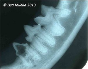
| − | |text = | + | |text = For interpretation dental radiographs should be viewed using a '''viewing box''' with minimal peripheral light and preferably using magnification. It is recommended to radiograph the '''contralateral structures for comparative purposes'''. |
|content = | |content = | ||
| − | :[[Normal Intra-Oral Radiographic Anatomy|Normal Radiographic Anatomy]] | + | :[[Normal Intra-Oral Radiographic Anatomy - Small Animal|Normal Radiographic Anatomy]] |
| − | :[[Dental Developmental Abnormalities - | + | :[[Radiographic Interpretation of Dental Developmental Abnormalities - Small Animal|Interpreting Developmental Abnormalities]] |
| − | :[[Periodontal Disease - | + | :[[Radiographic Interpretation of Periodontal Disease - Small Animal|Interpreting Periodontal Disease]] |
| − | :[[Endodontic Disease - | + | :[[Radiographic Interpretation of Endodontic Disease - Small Animal|Interpreting Endodontic Disease]] |
| − | :[[Tooth Resorption - | + | :[[Radiographic Interpretation of Tooth Resorption - Small Animal|Interpreting Tooth Resorption]] |
| − | :[[Dental Traumatic Injuries - | + | :[[Radiographic Interpretation of Dental Traumatic Injuries - Small Animal|Interpreting Traumatic Injuries]] |
| − | |image = | + | |image = Cat mandibular radiograph 1.jpg |
|resources = | |resources = | ||
}} | }} | ||
| + | |||
| + | {{Lisa Milella written | ||
| + | |date = 1 October 2014}} | ||
| + | |||
| + | {{Waltham}} | ||
| + | |||
| + | {{OpenPages}} | ||
[[Category:Intra-Oral Radiography]] | [[Category:Intra-Oral Radiography]] | ||
| + | [[Category:Waltham reviewed]] | ||
Latest revision as of 14:03, 2 November 2014
| ||||
|
| ||||
| This article was written by Lisa Milella BVSc DipEVDC MRCVS. Date reviewed: 1 October 2014 |
| Endorsed by WALTHAM®, a leading authority in companion animal nutrition and wellbeing for over 50 years and the science institute for Mars Petcare. |
Error in widget FBRecommend: unable to write file /var/www/wikivet.net/extensions/Widgets/compiled_templates/wrt6623c00c919af4_93996461 Error in widget google+: unable to write file /var/www/wikivet.net/extensions/Widgets/compiled_templates/wrt6623c00c964f71_19319428 Error in widget TwitterTweet: unable to write file /var/www/wikivet.net/extensions/Widgets/compiled_templates/wrt6623c00c998be7_66564768
|
| WikiVet® Introduction - Help WikiVet - Report a Problem |
