Leishmania
| This article is still under construction. |
| Leishmania spp. | |
|---|---|
| Kingdom | Eukaryota |
| Phylum | Euglenozoa |
| Class | Kinetoplastea |
| Order | Trypanosomatida |
| Family | Trypanosomatidae |
| Genus | Leishmania |
Overview
Leishmania spp. are intracellular parasites of macrophages from the same family as Trypanosoma spp.. These organisms parasitise human, dogs and wild animals throughout southern Europe, Africa, Asia and South America. The infection is transmitted by sandflies. Infection can cause both cutaneous and visceral disease. Three types of Leishmania spp. are described;
- Hypoplaria - found in lizards that ingest the sandfly intermediate host. Development occurs in the hindgut of the fly.
- Peripylaria - found in mammals and lizards, development occurs in the fore- and hindgut of the fly.
- Suprpylaria - found only in mammals transmitted by the bite of a sandfly, development occurs in the fore- and midgut of the fly.
Recognition
Leishmania spp. are ovoid shaped parasites containing a rod shaped 'kinetoplast'. The kinetoplast is associated with a rudimentary flagellum that does not extened beyond the cell margin. The position of the kinetoplast changes as the parasite changes between life stages. Once ingested by a sand fly the parasite takes the promastigote form and the kinetoplast moves the the posterior of the cell.
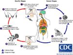
Life Cycle
- Transmitted by blood sucking sand flies
- Phlebotomus spp. in the Old World
- Lutzomyia spp. in the New World
- The amastigote (morphological form) is found in vertebrate macrophages
- Multiplies and migrates to insect proboscis
- Inoculated during feeding
- Can be transmitted percutaneously if sand fly crushed on skin
- Invades macrophages and reverts to amastigote
- Multiplies by binary fission
Pathogenesis
- Infection of vertebrate host
- Produces foci of proliferating Leishmania-infected macrophages in skin (cutaneous) or internal organs (visceral)
- Very long incubation period
- Months to years
- Many infected dogs are asymptomatic
- Visceral form causes chronic wasting condition
- Generalised eczema
- Loss of hair around eyes producing 'spectacle' effect
- Intermittent fever
- Generalised lymphadenopathy
- Generalised eczema
- Long periods of remission followed by recurrence of clinical signs is not uncommon in infections
- Involved in skin infections
Epidemiology
- Disease dependent on sand fly vectors
- E.g. Common in dogs around the Mediterranean coast, foci around southern Europe and around Madrid
- Reservoirs of infection
- E.g. Wild animals such as rodents and stray dogs
- Mechanisms of transmission
- sand fly bite
- Rarely through direct contact
- Leishmaniasis in British dogs
- Susceptible to infection if exposed whilst abroad in endemic areas as have no immunity
- No sand flies in Britain but dogs have become infected whilst in contact with infected imported animals
Diagnosis
- Demonstrate Leishmania organisms
- In skin scraping or smears
- In joint fluid, lymph node or bone marrow biopsies
Treatment and Control
- Chemotherapy
- Prolonged treatment, expensive, suppresses infection
- Does not cure infection
- Prevent sand flies biting
- Collars, sprays containing insecticide with repellent effect
- Destruction of infected and stray dogs
- Sand flies biting infected dogs may spread the disease to other dogs, humans and wildlife
- There is a slight possibility of transmission to humans by direct contact
In dogs
- disseminated disease of the monocyte-macrophage system
- protozoa; genus Leishmania
- increased travel means clinincal disease may be acquired in endemic areas but now presents to veterinarians in non-endemic areas
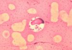
- lymph node aspirates contain macrophages with organisms


