Lungs - Anatomy & Physiology
Introduction


The lungs are the site for gaseous exchange, and are situated within the thoracic cavity. They occupy approximately 5% of the body volume in mammals when relaxed, and their elastic nature allows them to expand and contract with the processes of inspiration and expiration.
The lungs, along with the larynx and trachea, develop from a ventral respiratory tract. After separation from the developing oesophagus, two lung buds develop, which undergo divisions as they grow, forming the beginnings of the bronchial tree. This process is not completed at the time of parturition.
Structure
The left and right lungs lie within their pleural sac and are only attached by their roots, to the mediastinum, so they are fairly free within the thoracic cavity. The right lung is always larger than the left, due to the positioning of the heart. The apex of the lungs is their cranial point.
In most species, the lungs are divided into lobes by the bronchial tree:
Left Lung - Cranial and Caudal lobes.
Right Lung - Cranial, Caudal, Middle and Accessory lobes. The cranial lobe is further divided by an external fissure.
The bulk of the lung consists of bronchi, blood vessels and connective tissue. The terminal bronchioles have alveoli scattered along their length, and are continued by alveolar ducts, alveolar sacs and finally alveoli.
Alveolar Ducts
These have alveoli which open on all of its sides, they have no 'walls' as such. Openings to individual alveoli are guarded by smooth muscle.
Alveolar Sacs
These are rotunda-like areas on the end of each alveolar ducts which are usually present in clusters.
Alveoli
Alveoli are minute, polygonal chambers, whose diameter changes with the processes of inspiration and expiration, and varies by species. The wall of the alveoli is extremely thin, consisting of 2 irregular layers of epithelial sheets, 'sandwiching' a network of capillaries. Thus the Blood-Gas Barrier is just a single basal lamina - ideal for gaseous exchange. The alveolar interstitium is formed from connective tissue fibres and cells, which include collagen fibrils and elastin fibres.
Function
The main function of the lungs is gas exchange.
Vasculature
The pulmonary arteries follow the bronchi, while the pulmonary veins sometimes run separately. Bronchial arteries from the aorta supply the bronchi, and bronchial veins may drain this blood to the right atrium via the azygous vein. More often the blood from the bronchi drains directly to the left atrium.
Innervation
Nervous supply to the lung is via the pulmonary plexus within the mediastinum. The pulmonary plexus consists of sympathetic fibres largely from the stellate ganglion, and parasympathetic fibres from the vagus nerve.
Lymphatics
Lymph drains to the tracheobronchial and mediastinal lymph nodes.
Histology
Pneumocytes are a type of epithelial cells that lines the alveoli.
- the Type I pneumocytes are simple squamous with a flattened central nucleus that protrudes into the alveolar lume
- the Type II pneumocytes are round to pyramidal-shaped cells that are found among the type I pneumocytes. The type II pneumocytes have a larger centrally placed nucleus with a prominent nucleolus and a slightly vacuolated, foamy, basophilic cytoplasm. The nucleus of these cells also has a nucleolus.
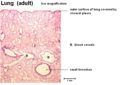
Lung (adult)
©RVC 2008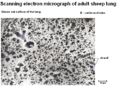
SEM of Adult Sheep Lung
©RVC 2008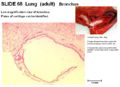
Bronchus
©RVC 2008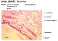
Bronchus
©RVC 2008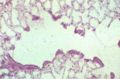
Alveoli
©RVC 2008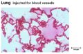
Alveoli
©RVC 2008


Species Differences
The lungs of the horse show almost no lobation, and the right lung of the horse lacks a middle lobe. In comparison to this, the lungs of ruminants and pigs are obviously lobed. The fissures between the lobes (interlobar fissures) are deeper in the dog and cat lung compared to other species. Avian respiration has many fundamental differences to mammalian respiration. The respiratory systems of non-homeotherms are also very different to that of mammals.
Links
Click here for more information on pathology of the lungs.
| Lungs - Anatomy & Physiology Learning Resources | |
|---|---|
Test your knowledge using drag and drop boxes |
Lung Histology Dragster |
 Selection of relevant videos |
Piglet Anatomy African Gray Parrot Radiograph Equine left-sided abdominal and thoracic topography dissection Equine thoracic cavity dissection Ovine right-sided abdominal and thoracic dissection |
 Selection of relevant PowerPoint tutorials |
Respiratory system histology tutorial with questions and answers |
Anatomy Museum Resources |
Dissection plans of the canine thoracic cavity Labelled dried dog lung Histology - Lung 1 Histology - Lung 2 Histology - Ovine Lung 1 Histology - Ovine Lung 2 Cast of Fallow deer lungs Interactive Pig Anatomy - Lungs |
References
Dyce, K.M., Sack, W.O. and Wensing, C.J.G. (2002) Textbook of Veterinary Anatomy. 3rd ed. Philadelphia: Saunders.
| This article has been peer reviewed but is awaiting expert review. If you would like to help with this, please see more information about expert reviewing. |
Error in widget FBRecommend: unable to write file /var/www/wikivet.net/extensions/Widgets/compiled_templates/wrt6621619c38b335_29448139 Error in widget google+: unable to write file /var/www/wikivet.net/extensions/Widgets/compiled_templates/wrt6621619c3bd000_05310442 Error in widget TwitterTweet: unable to write file /var/www/wikivet.net/extensions/Widgets/compiled_templates/wrt6621619c3eab53_08862997
|
| WikiVet® Introduction - Help WikiVet - Report a Problem |