Lymphangiectasia
| This article has been peer reviewed but is awaiting expert review. If you would like to help with this, please see more information about expert reviewing. |
Signalment
- Breed predisposition:
- Yorkshire Terrier
- Rottweiler
- Soft Coated Wheaten Terriers
- Lundehund
 Yorkshire Terrier
WikiCommons
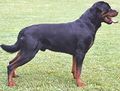 Rottweiler
WikiCommons
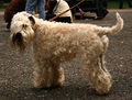 Soft Coated Wheaten Terrier
WikiCommons


Description
Lymphangiectasia is characterised by dilation and dysfunction of the lymphatic vessels of the intestines. Consequently, protein rich lymph leaks into the intestinal lumen, causing a protein-losing enteropathy and severe lipid malabsorption. It is relatively common in dogs but rare in cats.
Lymphangiectasia can be classified into a primary or a secondary lymphangiectasia. Primary lymphangiectasia may form part of a localised or a more widespread lymphatic abnormality. Secondary lymphangiectasia results from lymphatic obstruction, which may be caused by:
- inflammation,neoplastic infiltration or fibrosis
- thoracic duct obstruction
- right sided cardiac failure
- caval obstruction
- hepatic disease
Lymphangiectasia often accompanies a lipogranulomatous inflammation, but it is not clear which is the primary event. Lymphangitis can cause lymphatic obstruction but the leakage of lymph can also cause a granuloma to form.
Diagnosis
Clinical Signs
- Weight loss
- Chronic diarrhoea; steatorrhoea
- Ascites, oedema or chylothorax may result if there is severe hypoproteinaemia or lymphatic obstruction
- Increased appetite
- Vomiting, lethargy and anorexia (less common)
Laboratory Tests
Haematology
- Panhypoproteinaemia
- Lymphopaenia
Biochemistry
- Hypocholesterolaemia
- Hypocalcaemia due to hypoproteinaemia, vitamin D and calcium malabsorption
- Hypomagnesaemia

Other Tests
- Faecal α1-proteinase inhibitor concentrations or chromium 51-labelled albumin may be used to confirm protein-losing enteropathy.
Diagnostic Imaging
Ultrasound
Abdominal ultrasonography may reveal pleural fluid or ascites as well as helping to narrow down other differential diagnoses. Mucosa of intestinal loops may appear thickened due to oedema.
Endoscopy
Grossly, multiple white lipid droplets with prominent mucosal blebs can be seen.
Histopathology
Preferably, a full thickness biopsy is needed for a definitive diagnosis.
Refer to Lymphangiectasia for pathology
It is essential to distinguish a true lymphangiectasia from a secondary lacteal dilation due to Inflammatory Bowel Disease (IBD). In the case of IBD, inflammatory infiltrate will be seen in the lamina propria, but the degree of infiltration may be underestimated if oedema is present.
Treatment
- Identify and treat the underlying cause if it is a secondary lymphangiectasia
Dietary modification
- Fat-restricted diet
- The diet needs to be calorific and highly digestible
- Supplementation of fat soluble vitamins
- Anecdotal report of glutamine supplementation
Immunosuppressive
- Prednisolone
- Anti-inflammatory and immunosuppressive effect may be beneficial.
- This is particularly true if there is associated lymphangitis, lipogranulomas or a lymphocytic-plasmacytic infiltration of the lamina propria.
- Azathioprine or Ciclosporin can also be considered
Antimicrobials
- Metronidazole or tylosin can be given
- This may be beneficial due to their potential immunomodulatory effect and modulation of the enteric flora
- Diuretics such as frusemide and spironolactone are used to manage effusions.
Fluid therapy
Prognosis
Guarded. The response to treatment is generally poor although some dogs may do well. Dogs may be in remission for several years but the disease eventually progress to fulminant hypoproteinaemia.
From pathology
Pathology
Gross
- Small and large intestines may be affected.
- Dilation of lacteals.
- Oedema of intestinal mucosa.
- Distension of mesenteric lymphatics and lymph nodes.
Histological
- Accumulation of lipid-laden macrophages and granulomatous response around distended lymphatics.
References
- Ettinger, S.J. and Feldman, E. C. (2000) Textbook of Veterinary Internal Medicine Diseases of the Dog and Cat Volume 2 (Fifth Edition) W.B. Saunders Company.
- Hall, E.J, Simpson, J.W. and Williams, D.A. (2005) BSAVA Manual of Canine and Feline Gastroenterology (2nd Edition) BSAVA
- Nelson, R.W. and Couto, C.G. (2009) Small Animal Internal Medicine (Fourth Edition) Mosby Elsevier.