Myiasis
Also Known As – Fly-strike – Wound myiasis – Maggots – Strike – Fly infestation – Wool maggots
Caused By – Myiasis Producing Flies
Introduction
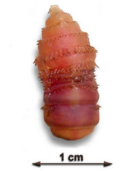
Myiasis is caused by infestation of live hosts with dipterous larvae.
Small and large ruminants and poultry can be affected.
These larvae then feed on the host’s living and dead tissue.
Myiasis Producing Flies can be obligate or facultative.
Myiasis Causing Flies
Obligate
Obligate flies occur exclusively in or on living vertebrates.
Oestrus ovis, Gasterophilus spp, Hypoderma spp, Dermatobia spp, Wohlfahrtia spp, Cochliomyia, Chrysomya bezziana and Cordylobia.
Facultative
Facultative flies are free-living and usually found in detritus or carrion.
Cochliomyia macellaria, Chrysomya megacephala, rufifacies and albiceps, Lucilla sericata and cuprina, Phormia spp, Protophormia spp and Caliphora spp.
For more information, see [Myiasis Producing Flies]]
Distribution
Myiasis Producing Flies are found in most regions of the world.
Signalment
Presence of wounds, wet fleece in sheep cases, recent surgery, bacterial wool/skin contamination and faecal contamination are the main predisposing factors for myiasis.
Density of stock will determine size and viability of the fly population.
Clinical Signs
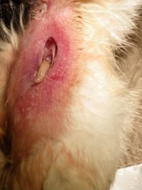
Can be classified as cutaneous, nasopharyngeal, intestinal or urogenital.
Pain, Irritation, Discomfort, Alopecia and Pruritus locally.
Direct tissue damage, haemorrhage, hyperpigmentation and secondary infection.
Nasal myiasis causes irritation and epistaxis.
Aural myiasis can cause deafness, discharge and foul exudates.
Gastrointestinal myiasis often causes ulceration, GI bleeding, weight loss, diarrhoea and pupae are voided in faeces.
Warble flies cause cysts along the midline of the back.
Lucilia sericata tends to cause leions on the inner thighs and perineum due to faecal soiling.
Wohlfahrtia spp cause genital lesions on the vulva and prepuce.
Loss of feathers and soiling of the vent is seen in poultry.
In severe cases, anaemia, anaphylaxis and toxaemia may be fatal.
Reduced feeding and consequent weight loss and infertility.
Diagnosis
Diagnosis is primarily dependent on observation of larvae on the host or in the faeces.
Larvae may also be observed in the carcass at post-mortem.
Gastroscopy may be used in the case of gastric myiasis.
ELISA is also available for Hypoderma spp and Oestrus ovis.
PCR is available for Cochliomyia spp.
Treatment
Treatment with ectoparasiticides is usually effective.
Administration can be oral, topical or by subcutaneous injection and the type of myiasis should be considered when deciding upon route of administration.
Ivermectin and doramectin are both effective in the control of nasal myiasis , warble fly and Dermatobia when injected.
Moxidectin is the main drug for oral treatment. It is effective against Gasterophilus spp.
Topical treatment by pour-on or dipping is most effective against cutaneous/subcutaneous myiasis. A huge range of products are available. Resistance should be considered and monitored.
Control
Preventative treatment with ectoparasiticides is common.
Release of sterile insects is also possible and effective but expensive.
Vaccines are available against Hypoderma spp and Lucilia spp.
References
Animal Health & ProductIon Compendium, Myiasis datasheet, accessed 06/06/2011 @ http://www.cabi.org/ahpc/

