Difference between revisions of "OVAM"
Ggaitskell (talk | contribs) |
Ggaitskell (talk | contribs) |
||
| Line 22: | Line 22: | ||
rect 0 0 900 900 [https://stream2.rvc.ac.uk/wikivet/Museum/rhino-pes1.wmv] | rect 0 0 900 900 [https://stream2.rvc.ac.uk/wikivet/Museum/rhino-pes1.wmv] | ||
desc none}} | desc none}} | ||
| − | <br><br><br> | + | <br><br><br><br> |
| + | |||
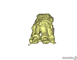
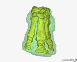
3D imaging of the bones in elephant and rhino by John Hutchinson, used to teach 3D skeletal anatomy. | 3D imaging of the bones in elephant and rhino by John Hutchinson, used to teach 3D skeletal anatomy. | ||
<br><br> | <br><br> | ||
| Line 29: | Line 30: | ||
desc none}} | desc none}} | ||
<br> | <br> | ||
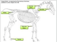
| − | Interactive drag and drop activity based on Stubbs work from 1766. | + | Interactive drag and drop activity based on Stubbs work from 1766. Accurate and detailed drawings based on dissection showing the full anatomy of the horse. |
<br><br> | <br><br> | ||
{{#tag:imagemap|File:Impact decimate.jpg{{!}}centre{{!}}200px | {{#tag:imagemap|File:Impact decimate.jpg{{!}}centre{{!}}200px | ||
| Line 39: | Line 40: | ||
desc none}} | desc none}} | ||
<br><br> | <br><br> | ||
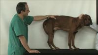
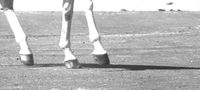
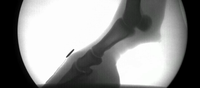
| − | Equine locomotion by Renate Weller | + | Equine locomotion by Renate Weller. |
|} | |} | ||
<!----------------------------------Manson Publishing-------------------------------> | <!----------------------------------Manson Publishing-------------------------------> | ||
Revision as of 14:51, 6 September 2011
The Online Veterinary Anatomy Museum (OVAM) will provide access to a comprehensive and pedagogically structured set of veterinary anatomical resources from UK veterinary schools and other institutions. These will be aggregated and ordered in an environment which will make them easily discoverable by different cohorts of learners. Key to the success of this project will be the development of effective methodologies to embed and integrate these materials within a traditional curriculum to maximise exposure, uptake and sustainability.

Partners
|
|
|
|