Difference between revisions of "OVAM"
Ggaitskell (talk | contribs) |
Ggaitskell (talk | contribs) |
||
| Line 44: | Line 44: | ||
|} | |} | ||
<br><br> | <br><br> | ||
| − | <!---------------------------------- | + | <!----------------------------------University of Murcia-------------------------------> |
|class="MainPageBG" style="width:50%; border:1px solid #cedff2; background:#f5faff; vertical-align:top;" colspan="1"| | |class="MainPageBG" style="width:50%; border:1px solid #cedff2; background:#f5faff; vertical-align:top;" colspan="1"| | ||
{|id="mp-right" width="100%" cellpadding="2" cellspacing="5" style="vertical-align:top; background:#f5faff;" | {|id="mp-right" width="100%" cellpadding="2" cellspacing="5" style="vertical-align:top; background:#f5faff;" | ||
| − | !<h2 style="margin:0; background:#cedff2; font-size:120%; font-weight:bold; border:1px solid #a3b0bf; text-align:left; color:#000; padding:0.2em 0.4em;">[ | + | !<h2 style="margin:0; background:#cedff2; font-size:120%; font-weight:bold; border:1px solid #a3b0bf; text-align:left; color:#000; padding:0.2em 0.4em;">[[Spain - Universidad de Murcia Facultad de Veterinaria]]</h2> |
|- | |- | ||
|style="color:#000;"| | |style="color:#000;"| | ||
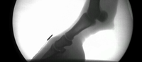
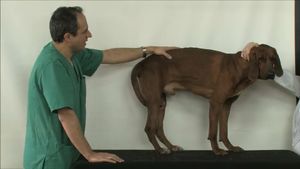
| − | {{#tag:imagemap|Image: | + | {{#tag:imagemap|Image:Abdomen video.jpg{{!}}centre{{!}}300px |
| − | rect 0 0 900 900 [http:// | + | rect 0 0 900 900 [http://www.um.es/anatvet/ingles/anatomy-videos.php] |
| + | desc none}} | ||
| + | <br> | ||
| + | Topographical anatomy videos by Octavio Lopez Albors. Visual learning aids showing the surface relationships and landmarks of the anatomy of the dog. | ||
| + | <br><br> | ||
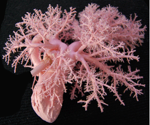
| + | {{#tag:imagemap|File:Plastination screenshot.png{{!}}centre{{!}}300px | ||
| + | rect 0 0 900 900 [http://www.um.es/museoveterinario/ingles/plastinated.php] | ||
desc none}} | desc none}} | ||
| − | + | <br> | |
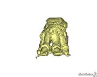
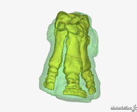
| + | Plastination of anatomy specimens. Preservation of dissection specimens using colored plastination techniques to show the routes of vasculature and nerves. | ||
|} | |} | ||
|} | |} | ||
| − | <!---------------------------------- | + | <!----------------------------------Manson Publishing-------------------------------> |
{|width="100%" style="margin:0px 0px 0px 0px; background:none;" | {|width="100%" style="margin:0px 0px 0px 0px; background:none;" | ||
|class="MainPageBG" style="width:50%; border:1px solid #cedff2; background:#f5faff; vertical-align:top;" colspan="1"| | |class="MainPageBG" style="width:50%; border:1px solid #cedff2; background:#f5faff; vertical-align:top;" colspan="1"| | ||
{|id="mp-right" width="100%" cellpadding="2" cellspacing="5" style="vertical-align:top; background:#f5faff;" | {|id="mp-right" width="100%" cellpadding="2" cellspacing="5" style="vertical-align:top; background:#f5faff;" | ||
| − | !<h2 style="margin:0; background:#cedff2; font-size:120%; font-weight:bold; border:1px solid #a3b0bf; text-align:left; color:#000; padding:0.2em 0.4em;">[ | + | !<h2 style="margin:0; background:#cedff2; font-size:120%; font-weight:bold; border:1px solid #a3b0bf; text-align:left; color:#000; padding:0.2em 0.4em;">[http://www.mansonpublishing.com/ Manson Publishing]</h2> |
|- | |- | ||
|style="color:#000;"| | |style="color:#000;"| | ||
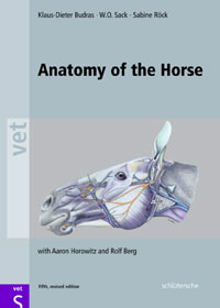
| − | {{#tag:imagemap|Image: | + | {{#tag:imagemap|Image:BudrasCover.jpg{{!}}centre{{!}}200px |
| − | rect 0 0 900 900 [http:// | + | rect 0 0 900 900 [http://upload.wikivet.net/images/f/fc/Budras_sample.pdf] |
desc none}} | desc none}} | ||
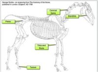
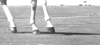
| − | + | A sample chapter from Budras' "Anatomy of the Horse" on the equine thoracic limb. Detailed information of all aspects of the anatomy of the equine limb, including skeletal, muscular, vascular and articular aspects, including descriptions and detailed diagrammatic drawings. | |
| − | + | ||
| − | |||
| − | |||
| − | |||
| − | |||
| − | |||
| − | |||
|} | |} | ||
<!----------------------------------Elsevier-------------------------------> | <!----------------------------------Elsevier-------------------------------> | ||
Revision as of 13:33, 7 September 2011
The Online Veterinary Anatomy Museum (OVAM) will provide access to a comprehensive and pedagogically structured set of veterinary anatomical resources from UK veterinary schools and other institutions. These will be aggregated and ordered in an environment which will make them easily discoverable by different cohorts of learners. Key to the success of this project will be the development of effective methodologies to embed and integrate these materials within a traditional curriculum to maximise exposure, uptake and sustainability.
|
|||||||||||||||||