Introduction
The Online Veterinary Anatomy Museum (OVAM) project was recently awarded funding by JISC. The official start of the project was the 1st November 2011 and it will run for 12 months.
The project will aim to provide access to veterinary anatomical resources in the form of a virtual museum. All UK vet schools are partners in the project as well as other European vet schools and commercial partners. The museum will be hosted within the WikiVet platform.
OVAM will provide access to a comprehensive and pedagogically structured set of veterinary anatomical resources from UK veterinary schools and other institutions. These will be aggregated and ordered in an environment which will make them easily discoverable by different cohorts of learners. Key to the success of this project will be the development of effective methodologies to embed and integrate these materials within a traditional curriculum to maximise exposure, uptake and sustainability.
For more information please see the OVAM Project Proposal Summary.
If you would be interested in collaboration with us, please do not hesitate to get in touch with Bara or Gemma
Visit our blog
Opening Event
The opening event for OVAM will be on the 9th December 2011 at the Royal Veterinary College's Hawkshead Campus. For more information please see the agenda. If you would like to attend please email Bara, places are limited.
| Anatomy Museum
|
Error in widget MediaCore: unable to write file /var/www/wikivet.net/extensions/Widgets/compiled_templates/wrt661e34409e6032_40051260
Partners
|
|
|
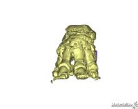
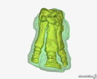
3D imaging of the bones in elephant and rhino by John Hutchinson, used to teach 3D skeletal anatomy.
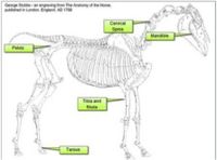
Interactive drag and drop activity based on Stubbs work from 1766. Accurate and detailed drawings based on dissection showing the full anatomy of the horse used to create interactive exercises for revision and learning.
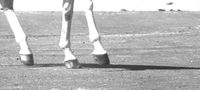
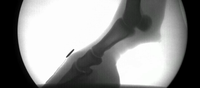
Equine locomotion by Renate Weller. Showing a horse trotting in slow motion focusing on the lower leg and the movement of joints through realtime radiography.
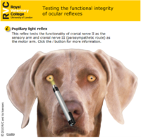
Interactive resource testing the functional integrity of the ocular reflex on a virtual patient in the form of a Flash animation. An example of a resource which helps to integrate anatomy teaching with the clinical years. Recently awarded first prize at JORUM, a national award for the best OER.
|
|
|
|
|
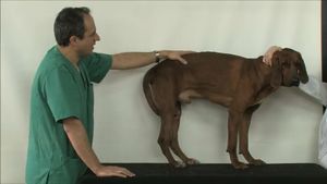
Topographical anatomy videos by Octavio Lopez Albors. Visual learning aids showing the surface relationships and landmarks of the anatomy of the dog.
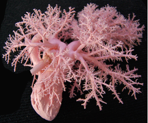
Plastination of anatomy specimens. Preservation of dissection specimens using colored plastination techniques to show the routes of vasculature and nerves.
|
|
|
|
|
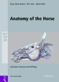
A sample chapter from Budras' "Anatomy of the Horse" on the equine thoracic limb. Detailed information of all aspects of the anatomy of the equine limb, including skeletal, muscular, vascular and articular aspects, including descriptions and detailed diagrammatic drawings.
|
|
|
|
|
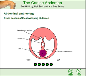
A sample from the Canine Abdomen by David Kilroy, Neil Stickland and Sue Evans of an introduction to abdominal embryology. Animations of the formation of the canine abdomen in cross section and accompanied by detailed descriptions.
|
|
Morpho Imaging - MOVE-IM
|
|
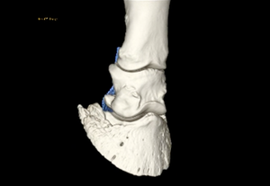
3D interactive multi-dimensional imaging of the equine foot by Dr Prisca Noble. Showing the bones, joint and position of tendons and other structures and pathologies of interest. Its objective is to teach a practical approach to veterinary anatomy that must be adapted by the clinician at work.
|
|
|
|
|
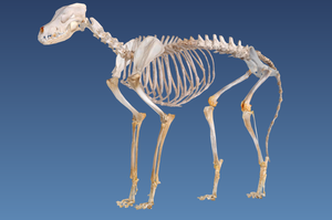
The canine skeleton in Real 3D Anatomy using anatomical models to aid e-learning, created using high quality detailed photographs, resulting in photorealistic 3D models. The presentations include hotspots, links and overlays.
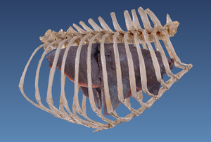
Canine thoracic organs in Real 3D Anatomy, showing the heart, lungs and ribs and allowing detailed and accurate visualisation of organs "in-situ", a valuable learning tool for anatomy students and clinical students studying approaches for surgical techniques.
|
|
|
|
|
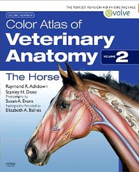
Sample drag and drop exercises from Ashdowns' Color Atlas of Veterinary Anatomy: The Horse. Labels have to be dragged to the correct position on photographs, anatomical diagrams and radiographs. A useful tool for revision and reinforcement of learning.
|
|
|
|
|
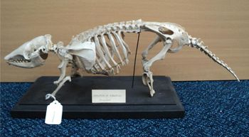
Skeletal anatomy of the Armadillo allowing comparison of the anatomy of wild and domestic species.
|
|
|
|
|

|
This project was funded by the Jisc Content Programme for 2011-13.
Jisc inspires UK colleges and universities in the innovative use of digital technologies, helping to maintain the UK's position as a leader in education and research.
|