Ophidian Paramyxovirus
| This article is still under construction. |
Species predisposition
Ophidian paramyxovirus (OPMV), also called Fer-de-Lance virus of reptiles (FDLV), is an important viral pathogen of snakes, principally Viperidae and it has also been isolated from Elapidae, Colubridae and Boidae. The natural host for OPMV is unknown but may be a non-viperid. Disease may be associated with decreased immunocompetence. Transmission may be by droplet, fomites and vectors such as mites. Congenital infection may be involved.
Examination
Anorexia may be seen several weeks before presentation. Clinical signs are often related to the respiratory system. Presentation is frequently a moribund snake. There may be a discharge from the glottis that is sometimes bloody (haemorrhagic pneumonia). Central nervous system signs (writhing, torticollis, loss of righting reflex) are occasionally seen.
- For more information, see snake physical examination.
Diagnosis
OPMV should be suspected in Viperidae presented with signs associated with immunodeficiency. There are several methods of diagnosis.
Antemortem
A haemaglutination inhibition test (HI) for specific antibodies to OPMV has been developed (titre < 1:20 - negative, 1:40 to 1:80 - suspect and > 1:80 - positive). A positive titre means exposure and not disease or shedding status. Two samples for HI at a 2-4 week interval are advisable. Negative staining electron microscopy of faecal or lung samples is possible. A polymerase chain reaction (PCR) would be very useful.
Postmortem
- Gross necropsy - Lesions are usually subtle to non-existent but may include pulmonary (pneumonia - congestion, oedema, haemorrhage or inflammatory exudate) and pancreatic (necrosis, hyperplasia, interstitial oedema or fibrosis) lesions. The liver may be involved.
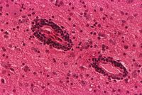
- Histopathology - There may be characteristic histopathological lesions in lung and pancreas and occasionally CNS. Sections from cranial, mid and caudal lung should be examined. Immunoperoxidase and immunofluorescent staining is possible (formalined-fix tissues for 48 hours then store in 70% ethanol).
- Virus isolation is the definitive diagnostic tool. Tissue (lung, liver, kidney, spleen and heart) is placed in individual sterile bage and frozen.
Therapy
There is no specific treatment for snakes showing clinical signs of OPMV infection. Antibiotics are indicated because most affected snakes die with gram-negative respiratory tract infections.
- See also snake respiratory disease.
Prevention
The best control is a sound preventative medicine programme. All new specimens should be quarantined for ninety days. Hygiene is very important because of the possibility of spread by fomites and mites. Currently, there is no vaccine available for protecting snakes against OPMV.
