Difference between revisions of "Oral Examination Under General Anaesthesia"
Ggaitskell (talk | contribs) |
|||
| (3 intermediate revisions by the same user not shown) | |||
| Line 1: | Line 1: | ||
| + | {{OpenPagesTop}} | ||
==Introduction== | ==Introduction== | ||
The endotracheal tube does not allow full closure of the mouth to examine the relationship between the teeth. In the anaesthetised patient prior to intubation, the muscles are relaxed and the tongue often gets in the way, so a complete evaluation of occlusion is not always possible. | The endotracheal tube does not allow full closure of the mouth to examine the relationship between the teeth. In the anaesthetised patient prior to intubation, the muscles are relaxed and the tongue often gets in the way, so a complete evaluation of occlusion is not always possible. | ||
| Line 85: | Line 86: | ||
#Mobility | #Mobility | ||
| − | {{ | + | |
| + | {{Lisa Milella written | ||
| + | |date = 13 August 2014}} | ||
| + | |||
| + | {{Waltham}} | ||
| + | |||
| + | {{OpenPages}} | ||
[[Category:Oral Examination]] | [[Category:Oral Examination]] | ||
| − | [[Category: | + | [[Category:Waltham reviewed]] |
Latest revision as of 14:09, 2 November 2014
Introduction
The endotracheal tube does not allow full closure of the mouth to examine the relationship between the teeth. In the anaesthetised patient prior to intubation, the muscles are relaxed and the tongue often gets in the way, so a complete evaluation of occlusion is not always possible.
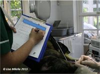
The oropharynx should be examined prior to endotracheal intubation. Normal anatomical features of the oral cavity need to be identified and inspected. A check list is given below:
|
|
|
|
|

Any abnormalities need to be noted – look for swellings, inflammation, ulcerations. Check if the lesion is localised to one area or more generalised. Always biopsy abnormal tissue if a cause cannot be identified.
Under general anaesthesia, it is also useful to recheck the temporomandibular joints for crepitus or clicks if a problem is suspected. The mandibular symphysis should also be checked for mobility – a small degree of movement is normal in cats.
Indices and Criteria
The following indices and criteria should be evaluated for each tooth:
- Gingivitis and gingival index
- Periodontal probing depth
- Gingival recession
- Furcation involvement
- Mobility
| This article was written by Lisa Milella BVSc DipEVDC MRCVS. Date reviewed: 13 August 2014 |
| Endorsed by WALTHAM®, a leading authority in companion animal nutrition and wellbeing for over 50 years and the science institute for Mars Petcare. |
Error in widget FBRecommend: unable to write file /var/www/wikivet.net/extensions/Widgets/compiled_templates/wrt6621a6e8bf07f0_90381714 Error in widget google+: unable to write file /var/www/wikivet.net/extensions/Widgets/compiled_templates/wrt6621a6e8c774d7_97951850 Error in widget TwitterTweet: unable to write file /var/www/wikivet.net/extensions/Widgets/compiled_templates/wrt6621a6e8cbd4e8_40862080
|
| WikiVet® Introduction - Help WikiVet - Report a Problem |