Radiographic Interpretation of Dental Developmental Abnormalities - Small Animal
Developmental Abnormalities
Introduction
Dental developmental abnormalities occur commonly in dogs and occasionally in cats. They can be caused by abnormal genetic coding or by damage to developing tissues. The necessity for treatment is based on whether the abnormality negatively impacts the health, function, or comfort of the patient. Radiographic evaluation of the extent of involvement helps the clinician to determine which abnormalities require immediate intervention, which might require treatment or monitoring, and which do not require any intervention at all.
Abnormal Tooth Shape or Structure
- Enamel dysplasia can be caused by systemic diseases (for example, viral infection such as distemper) or trauma during development, and also by hereditary or nutritional factors. Radiographs should be made to determine whether tooth root development was also affected.
- Supernumerary roots most commonly affect maxillary third premolar teeth in both dogs and cats. Most are not associated with pathology and may be considered a variation of normal. These can be clinically significant if associated with periodontitis, and during extraction or endodontic treatment of the tooth.
- Fusion of two tooth germs results in a single large tooth and one fewer tooth in the arch.
- Gemination is the attempt of a single enamel organ to make two teeth. The result is a tooth with two crowns on one root. In single-rooted teeth, this cannot be differentiated from the fusion of the root of a normal tooth with that of a supernumerary tooth. In multirooted teeth, fusion of supernumerary teeth is more apparent. When complete separation forms two teeth from one tooth germ, the result is a type of supernumerary tooth called “twinning”. Supernumerary mandibular fourth premolar teeth are commonly bilateral in cats.
- Convergent roots occur most commonly on mandibular first molar teeth in small-breed dogs. Affected teeth frequently develop endodontic disease. The clinical crowns of affected molar teeth often appear normal except for a developmental groove close to the mid-buccal gingival margin. Premolar teeth can also have convergent roots, sometimes with incomplete separation of the roots during development.
- Dens in dente, or dens invaginatus, is an infolding of the developing tooth of varying severity that can result in periodontal problems and endodontic involvement.
- Enamel pearls are deposits of enamel on the root that sometimes have dentin involvement and rarely pulp as well. As the periodontal ligament is unable attach to enamel they may contribute to periodontitis.
Abnormal Tooth Number
Hypodontia, or missing teeth, usually presents no clinical problems for dogs and cats but the missing tooth area should be radiographed to confirm that the tooth is truly missing and not unerupted or impacted, which could potentially lead to complications.
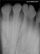
Fused incisors
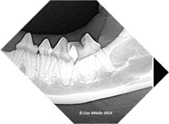
Supernumerary teeth
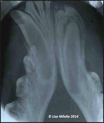
Impacted tooth


| Radiographic Interpretation of Dental Developmental Abnormalities - Small Animal Learning Resources | |
|---|---|
To reach the Vetstream content, please select |
Canis, Felis, Lapis or Equis |
| This article was written by Lisa Milella BVSc DipEVDC MRCVS. Date reviewed: 13 August 2014 |
| Endorsed by WALTHAM®, a leading authority in companion animal nutrition and wellbeing for over 50 years and the science institute for Mars Petcare. |
Error in widget FBRecommend: unable to write file /var/www/wikivet.net/extensions/Widgets/compiled_templates/wrt662287d3321395_57002579 Error in widget google+: unable to write file /var/www/wikivet.net/extensions/Widgets/compiled_templates/wrt662287d335b495_16101439 Error in widget TwitterTweet: unable to write file /var/www/wikivet.net/extensions/Widgets/compiled_templates/wrt662287d3390121_05027192
|
| WikiVet® Introduction - Help WikiVet - Report a Problem |