Difference between revisions of "Radiographic Interpretation of Periodontal Disease - Small Animal"
| (5 intermediate revisions by the same user not shown) | |||
| Line 1: | Line 1: | ||
| + | {{OpenPagesTop}} | ||
==Interpreting Periodontal Disease== | ==Interpreting Periodontal Disease== | ||
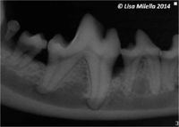
[[File:Alveolar bone destruction.jpg|right|200px|thumb|Alveolar bone destruction]] | [[File:Alveolar bone destruction.jpg|right|200px|thumb|Alveolar bone destruction]] | ||
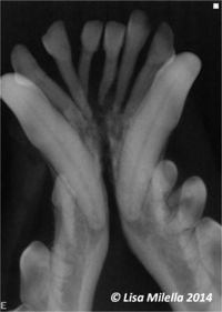
| − | [[Intra-Oral Radiography - Small Animal|Dental radiographs]] assist in the assessment of [[Periodontal Disease|periodontitis]] by providing information regarding [[ | + | [[File:Severe periodontitis (grade 4).jpg|200px|right|thumb|[[Periodontal Disease#Diagnosis|Grade 4 periodontitis]]]] |
| + | [[Intra-Oral Radiography - Small Animal|Dental radiographs]] assist in the assessment of '''[[Periodontal Disease|periodontitis]] '''by providing information regarding [[Tooth - Anatomy & Physiology#Alveolar Bone|alveolar bone]] loss. They complement, but do not replace, the [[Oral Examination - Small Animal|clinical examination]]. Clinical examination is essential for evaluating soft tissue changes such as inflammation, [[Dental Indices and Criteria#Gingival Recession|gingival recession]], and periodontal pocket formation. Clinical examination will provide evidence of mild bone loss, such as a Grade I [[Dental Indices and Criteria##Furcation Involvement|furcation exposure]], prior to changes being apparent on a dental radiograph. The dental radiograph is a two-dimensional image, and the morphology of an infrabony defect will be determined on clinical examination rather than on radiographic evaluation. | ||
<br><br> | <br><br> | ||
| − | Widening of the [[ | + | Widening of the [[Tooth - Anatomy & Physiology#Periodonal Ligament|periodontal ligament]] space, decreased alveolar bone density, and bone loss are all radiographic changes associated with periodontitis.<br><br> |
Terms used to describe the alveolar bone loss associated with periodontitis include: | Terms used to describe the alveolar bone loss associated with periodontitis include: | ||
*Alveolar margin bone loss | *Alveolar margin bone loss | ||
| Line 16: | Line 18: | ||
<br><br> | <br><br> | ||
| + | |||
| + | {{Lisa Milella written | ||
| + | |date = 10 April 2014}} | ||
| + | |||
| + | {{Waltham}} | ||
| + | |||
| + | {{OpenPages}} | ||
[[Category:Intra-Oral Radiography]] | [[Category:Intra-Oral Radiography]] | ||
[[Category:Periodontal Conditions]] | [[Category:Periodontal Conditions]] | ||
| − | [[Category: | + | [[Category:Waltham reviewed]] |
Latest revision as of 14:16, 2 November 2014
Interpreting Periodontal Disease
Dental radiographs assist in the assessment of periodontitis by providing information regarding alveolar bone loss. They complement, but do not replace, the clinical examination. Clinical examination is essential for evaluating soft tissue changes such as inflammation, gingival recession, and periodontal pocket formation. Clinical examination will provide evidence of mild bone loss, such as a Grade I furcation exposure, prior to changes being apparent on a dental radiograph. The dental radiograph is a two-dimensional image, and the morphology of an infrabony defect will be determined on clinical examination rather than on radiographic evaluation.
Widening of the periodontal ligament space, decreased alveolar bone density, and bone loss are all radiographic changes associated with periodontitis.
Terms used to describe the alveolar bone loss associated with periodontitis include:
- Alveolar margin bone loss
- Furcation bone loss
- Horizontal bone loss
- Vertical bone loss
- A combination of horizontal and vertical bone loss
Dental radiographs made for the evaluation of early alveolar bone loss should not be overexposed as this may result in “burn-out” of the alveolar marginal and interdental marginal bone. Dental radiographs should be made at the correct exposure or should be slightly under-exposed to provide the highest level of detail when evaluating for early bone loss.
Alveolar bone loss may be mild, moderate, or severe. The significance of the bone loss will depend on the amount of attachment loss and the teeth involved. Furcation bone loss is bone loss that occurs in the area where multirooted teeth divide. Approximately 30% to 40% of the furcation bone must be lost before it is evident on a radiograph. A Grade 3 furcation exposure is complete loss of bone in the furcation area, and this may be identified readily on most two-rooted teeth but may be more difficult to determine on the three-rooted maxillary molar teeth.
Crowded and malpositioned teeth make it more difficult to diagnose alveolar margin and furcation bone loss from a radiograph. Interdental alveolar bone loss and furcation bone loss, especially when mild, are often easier to identify radiographically than alveolar bone loss superimposed over the tooth roots.
Severe periodontitis may result in secondary complications such as external root resorption and endodontic disease or weakening of the mandible and therefore potential for fracture.
| This article was written by Lisa Milella BVSc DipEVDC MRCVS. Date reviewed: 10 April 2014 |
| Endorsed by WALTHAM®, a leading authority in companion animal nutrition and wellbeing for over 50 years and the science institute for Mars Petcare. |
Error in widget FBRecommend: unable to write file /var/www/wikivet.net/extensions/Widgets/compiled_templates/wrt6621b0b41a7b34_23906162 Error in widget google+: unable to write file /var/www/wikivet.net/extensions/Widgets/compiled_templates/wrt6621b0b4200143_23790331 Error in widget TwitterTweet: unable to write file /var/www/wikivet.net/extensions/Widgets/compiled_templates/wrt6621b0b4239074_60065381
|
| WikiVet® Introduction - Help WikiVet - Report a Problem |

