Difference between revisions of "Snake Physical Examination"
| Line 3: | Line 3: | ||
==Introduction== | ==Introduction== | ||
The physical examination involves observation of the snake, taking measurements and a thorough methodical area by area examination. Many techniques are similar to other animals, but before examining the snake ask the owner if it is accustomed to being handled. [[Snake Handling and Restraint|See here]] for information on handling and restraint of snakes. A veterinarian who is inexperienced with reptiles may be likely to focus on the presenting signs but then can end up treating exclusively the secondary problems. Stomatitis and rectal prolapse are secondary conditions where a full examination with husbandry review, including [[Snake Housing|housing]] and [[Snake feeding|nutrition]], is vital in determining the principal problem. | The physical examination involves observation of the snake, taking measurements and a thorough methodical area by area examination. Many techniques are similar to other animals, but before examining the snake ask the owner if it is accustomed to being handled. [[Snake Handling and Restraint|See here]] for information on handling and restraint of snakes. A veterinarian who is inexperienced with reptiles may be likely to focus on the presenting signs but then can end up treating exclusively the secondary problems. Stomatitis and rectal prolapse are secondary conditions where a full examination with husbandry review, including [[Snake Housing|housing]] and [[Snake feeding|nutrition]], is vital in determining the principal problem. | ||
| − | + | ==Observation== | |
| − | Snakes should have active tongues that are sampling scent particles in the atmosphere. Normal movement should be observed although allowances must be made for any chilling effect in transit since this will reduce the patient's metabolism and give a misleading impression of lethargy and lack of strength. The righting reflex should be tested since poor reactions can be a result of weakness and not necessarily neurological disease. Respiration, including rib movements and any sounds produced | + | Snakes should have active tongues that are sampling scent particles in the atmosphere. Normal movement should be observed although allowances must be made for any chilling effect in transit since this will reduce the patient's metabolism and give a misleading impression of lethargy and lack of strength. A healthy snake will have adequate muscle tone and grip the clinician's hands. There will be a level of activity befitting the particular species. A sick snake usually remains limp. The righting reflex should be tested since poor reactions can be a result of weakness and not necessarily neurological disease. Neurological function can be assessed by checking head position, body posture, cloacal tone and righting reflexes. Respiration, including rib movements and any sounds produced should be noted. |
| − | == | + | ==Organ positions== |
| − | + | The organisation of internal structures varies between snake families and genera. However it is important to know the approximate position of the organs during a physical examination. The following is a generalisation if the snake's body is divided into thirds from snout to vent: | |
| + | *'''Cranial third''' - contains the trachea, oesophagus and heart | ||
| + | *'''Middle third''' - contains the liver, lung and stomach | ||
| + | *'''Caudal third''' - contains the small intestine, the triad of pancreas, spleen and gallbladder, the adrenal glands, large intestine, gonads, reproductive tract, kidneys, fat body and cloaca. Behind the vent are the musk and, if the snake is a male, the hemipenes. | ||
==Skin== | ==Skin== | ||
[[Image:0051_BLISTER_DISEASE_PYTHON_ed.jpg|250px|thumb|right|'''Blisters on skin''' ©RVC and its licensors, Peer Zwart and Fredric Frye. All rights reserved]] | [[Image:0051_BLISTER_DISEASE_PYTHON_ed.jpg|250px|thumb|right|'''Blisters on skin''' ©RVC and its licensors, Peer Zwart and Fredric Frye. All rights reserved]] | ||
| − | + | Check the general appearance of the skin. The skin should be bright and shiny. Assess elasticity and skin tenting. Dehydrated snakes show outwardly directed folds of skin and dysecdysis. Examine the skin both dorsally and ventrally for retained shed, blisters, pustules, burns, discharges, erythema, pettechi, ulcers, loss of scales, lacerations ad swellings. Examine for ectoparasites, especially mites that tend to hde under scales of the skin. Brushing the body of the snake over a white sheet may be helpful in checking for mites. The snake's body can be palpated by simply moving the hand, both dorsally and ventrally, down its length. Identify any masses and swellings under the skin. Subcutaneous lumps are usually due to abscesses but neoplasia and the second-stage cysts of cestodes can also be encountered in snakes. | |
| − | |||
| − | |||
| − | |||
| − | |||
| − | |||
| − | |||
| − | |||
| − | |||
| − | |||
| − | |||
| − | |||
Find out more about [[Snake Skin|snake skin]] | Find out more about [[Snake Skin|snake skin]] | ||
| Line 29: | Line 21: | ||
Ecdysis (shedding) generally takes place every 14 days although this is dependent on many variables such as environment, age and size of snake. Snakes shed their entire skin in one piece, including the spectacles, although large snakes greater than 3m in length may shed their skin in incomplete sections. Prior to ecdydsis, a snake will become anorectic and handling may be hazardous to the animal at this time if the underlying epidermis is damaged. | Ecdysis (shedding) generally takes place every 14 days although this is dependent on many variables such as environment, age and size of snake. Snakes shed their entire skin in one piece, including the spectacles, although large snakes greater than 3m in length may shed their skin in incomplete sections. Prior to ecdydsis, a snake will become anorectic and handling may be hazardous to the animal at this time if the underlying epidermis is damaged. | ||
==Head== | ==Head== | ||
| − | + | Initially check that the snake moves and supports its head normally and that it investigates its surroundings with an active flicking tonge. Examine the rostrum for abrasions that may indicate an unsuitable cage. Check the nares for patency and discharge. Examine the heat-sensing pits, the crevice around the spectacle and the skin between the scales for ectoparasites or retained skin. Palpate the jaws for any abnormal swellings. | |
| − | |||
===Eyes=== | ===Eyes=== | ||
| − | The eyes are usually considered as a barometer of general health and environmental conditions; therefore a full ophthalmologic examination should be performed in all cases. | + | The eyes are usually considered as a barometer of general health and environmental conditions; therefore a full ophthalmologic examination should be performed in all cases. Observe the movement and general position of the eye within the socket. The optic nerve and other cranial nerves associated with eye function are assessed by observing the snake's reaction to movement and by normal and co-ordinated eye movements. Strabismus may indicated dysfunction. Bulging eyes may result from retrobulbar or periocular cellulitis, absecesses or neoplasia. Inspect the spectacles. They should be clear, not wrinkled, distended or discoloured although they are normally bluish-white prior to shedding. The subspectacular space should be clear. Check for any signs of conjunctivitis. Remember that the mydriatics are not useful and that there is no consensual light reflex. |
| − | + | * Other diagnostic tools, such as tonometry (measurement of the intraocular pressure), stains, cytology, bacteriology, histopathology and electron microscopy, in addition to routine diagnostic tools (haemotology, biochemistry, radiology and ultrasound) can also be used to detect an ocular disease or underlying problem in snakes. | |
| − | |||
| − | |||
| − | |||
| − | * Other diagnostic tools, such as tonometry (measurement of the intraocular pressure), stains, cytology, bacteriology, histopathology and electron microscopy, in addition to routine diagnostic tools (haemotology, biochemistry, radiology and ultrasound) can also be used to detect an ocular disease or underlying problem in snakes | ||
| − | |||
[[Image:Boa_stomatitis_ed.jpg|200px|thumb|right|'''Ulcerative stomatitis''' ©RVC and its licensors, Peer Zwart and Fredric Frye. All rights reserved]] | [[Image:Boa_stomatitis_ed.jpg|200px|thumb|right|'''Ulcerative stomatitis''' ©RVC and its licensors, Peer Zwart and Fredric Frye. All rights reserved]] | ||
===Oral cavity=== | ===Oral cavity=== | ||
| − | + | The oral cavity is fairly easy to evaluate once the mouth is open. Use an object to gently open the mouth without damaging the delicate gingiva and teeth. Do not use any cotton since it snags on the caudally curving teeth. The normally pale and pinkish-white gingiva should be glistening and moist with no petecchiation, excess salivation or oedema. Excessive saliva in the oral cavity suggests pneumonia or gastrointestinal upset. Mouth rot when present varies from mild erythema to a generalised stomatitis with pus necrotic tissue. The tongue and its sheath should be examined. Check the glottis for any discharge. Check the patency of the internal nares. | |
| − | + | ==Musculoskeletal== | |
| − | + | Assess the bulk and tone of the muscles first. The body should be rounded. With emaciation it becomes triangular and the spine and ribs more prominent. Then assess locomotion on a rough surface. Normal posture and coordination should be evident. Check the righting reflex. A defect in the vestibular system or hypothiaminosis may result in rolling and an inability to right. The spine and ribcage should then be palpated for swellings that may be associated with abscesses or fractures. | |
| − | + | ==Auscultation/cardiovascular== | |
| − | + | In colubrids and boids the heart is located in the cranial third of the body and the lung is in the middle third. The use of a moistened gauze sponge over the stethoscope will help to eliminated the grating noise against the scales. Special attention should be paid to abnormal sounds and symmetry. Sometimes the presence of mucous can be detected. Defensive hissing must be differentiated from pathologic respiration. | |
| − | + | ==Coloemic cavity== | |
| + | Examination of the coelomic cavity is possible in most small snakes by palpating ventrally between the ends of the ribs. Large snakes are difficult to palpate because of their musculature. There should be no palpable swellings and if they do occur their position can indicate the organ involved. The heart can be palpated in the cranial third and the liver lies at a distance caudally in boids and colubrids because of their caudal lung. Caudal to the liver the gall bladder can be palpated on the ventral midline. It is possible to palpate the ingesta in healthy snakes while anorexic individuals will feel empty of doughy. | ||
| + | ==Vent and tail== | ||
| + | The cloaca can be examined very simply by using a otoscope or endoscope. Check for oedema, erythema, discharge and swellings. Check the vent for any soiling which may be due to parasitic infection of the large intestine. Make a faecal smear if possible. Examine the tail for retained sloughed skin, lumps and other deformities. | ||
| + | [[Image:Sexing_snakes.jpg|200px|thumb|right|'''Sexing snakes''' ©RVC and its licensors, Peer Zwart and Fredric Frye. All rights reserved]] | ||
| + | ==Sexing/Probing== | ||
| + | The snake is positioned so that the vent is easily accessible. A smooth lubricated blunt-tipped probe is inserted caudally through the vent. In males the probes enters the hemipenes (10-12 scale rows) while in females it may enter the musk glands (2-3 scale rows). Take care since rough use of probes can cause damage. In females it is possible to perforate the musk gland and allow the probe to slide in as far as it would in a male snake. | ||
==References== | ==References== | ||
Fowler, M.E. and Miller, R.E. (2003). Zoo and Wild Animal Medicine. Saunders, 5th Edition. pp. 84. ISBN 0-7216-9499-3 | Fowler, M.E. and Miller, R.E. (2003). Zoo and Wild Animal Medicine. Saunders, 5th Edition. pp. 84. ISBN 0-7216-9499-3 | ||
[[Category:Snake_Examination]] | [[Category:Snake_Examination]] | ||
Revision as of 17:55, 14 March 2010
| This article is still under construction. |
Introduction
The physical examination involves observation of the snake, taking measurements and a thorough methodical area by area examination. Many techniques are similar to other animals, but before examining the snake ask the owner if it is accustomed to being handled. See here for information on handling and restraint of snakes. A veterinarian who is inexperienced with reptiles may be likely to focus on the presenting signs but then can end up treating exclusively the secondary problems. Stomatitis and rectal prolapse are secondary conditions where a full examination with husbandry review, including housing and nutrition, is vital in determining the principal problem.
Observation
Snakes should have active tongues that are sampling scent particles in the atmosphere. Normal movement should be observed although allowances must be made for any chilling effect in transit since this will reduce the patient's metabolism and give a misleading impression of lethargy and lack of strength. A healthy snake will have adequate muscle tone and grip the clinician's hands. There will be a level of activity befitting the particular species. A sick snake usually remains limp. The righting reflex should be tested since poor reactions can be a result of weakness and not necessarily neurological disease. Neurological function can be assessed by checking head position, body posture, cloacal tone and righting reflexes. Respiration, including rib movements and any sounds produced should be noted.
Organ positions
The organisation of internal structures varies between snake families and genera. However it is important to know the approximate position of the organs during a physical examination. The following is a generalisation if the snake's body is divided into thirds from snout to vent:
- Cranial third - contains the trachea, oesophagus and heart
- Middle third - contains the liver, lung and stomach
- Caudal third - contains the small intestine, the triad of pancreas, spleen and gallbladder, the adrenal glands, large intestine, gonads, reproductive tract, kidneys, fat body and cloaca. Behind the vent are the musk and, if the snake is a male, the hemipenes.
Skin
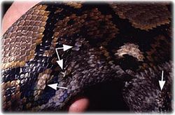
Check the general appearance of the skin. The skin should be bright and shiny. Assess elasticity and skin tenting. Dehydrated snakes show outwardly directed folds of skin and dysecdysis. Examine the skin both dorsally and ventrally for retained shed, blisters, pustules, burns, discharges, erythema, pettechi, ulcers, loss of scales, lacerations ad swellings. Examine for ectoparasites, especially mites that tend to hde under scales of the skin. Brushing the body of the snake over a white sheet may be helpful in checking for mites. The snake's body can be palpated by simply moving the hand, both dorsally and ventrally, down its length. Identify any masses and swellings under the skin. Subcutaneous lumps are usually due to abscesses but neoplasia and the second-stage cysts of cestodes can also be encountered in snakes.
Find out more about snake skin
Find out more about snake skin diseases
Shedding
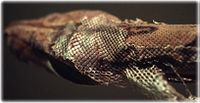
Ecdysis (shedding) generally takes place every 14 days although this is dependent on many variables such as environment, age and size of snake. Snakes shed their entire skin in one piece, including the spectacles, although large snakes greater than 3m in length may shed their skin in incomplete sections. Prior to ecdydsis, a snake will become anorectic and handling may be hazardous to the animal at this time if the underlying epidermis is damaged.
Head
Initially check that the snake moves and supports its head normally and that it investigates its surroundings with an active flicking tonge. Examine the rostrum for abrasions that may indicate an unsuitable cage. Check the nares for patency and discharge. Examine the heat-sensing pits, the crevice around the spectacle and the skin between the scales for ectoparasites or retained skin. Palpate the jaws for any abnormal swellings.
Eyes
The eyes are usually considered as a barometer of general health and environmental conditions; therefore a full ophthalmologic examination should be performed in all cases. Observe the movement and general position of the eye within the socket. The optic nerve and other cranial nerves associated with eye function are assessed by observing the snake's reaction to movement and by normal and co-ordinated eye movements. Strabismus may indicated dysfunction. Bulging eyes may result from retrobulbar or periocular cellulitis, absecesses or neoplasia. Inspect the spectacles. They should be clear, not wrinkled, distended or discoloured although they are normally bluish-white prior to shedding. The subspectacular space should be clear. Check for any signs of conjunctivitis. Remember that the mydriatics are not useful and that there is no consensual light reflex.
- Other diagnostic tools, such as tonometry (measurement of the intraocular pressure), stains, cytology, bacteriology, histopathology and electron microscopy, in addition to routine diagnostic tools (haemotology, biochemistry, radiology and ultrasound) can also be used to detect an ocular disease or underlying problem in snakes.
Oral cavity
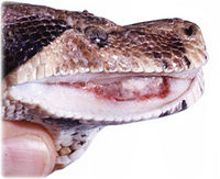
The oral cavity is fairly easy to evaluate once the mouth is open. Use an object to gently open the mouth without damaging the delicate gingiva and teeth. Do not use any cotton since it snags on the caudally curving teeth. The normally pale and pinkish-white gingiva should be glistening and moist with no petecchiation, excess salivation or oedema. Excessive saliva in the oral cavity suggests pneumonia or gastrointestinal upset. Mouth rot when present varies from mild erythema to a generalised stomatitis with pus necrotic tissue. The tongue and its sheath should be examined. Check the glottis for any discharge. Check the patency of the internal nares.
Musculoskeletal
Assess the bulk and tone of the muscles first. The body should be rounded. With emaciation it becomes triangular and the spine and ribs more prominent. Then assess locomotion on a rough surface. Normal posture and coordination should be evident. Check the righting reflex. A defect in the vestibular system or hypothiaminosis may result in rolling and an inability to right. The spine and ribcage should then be palpated for swellings that may be associated with abscesses or fractures.
Auscultation/cardiovascular
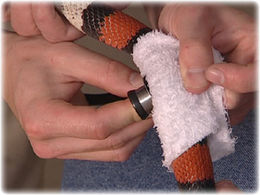
In colubrids and boids the heart is located in the cranial third of the body and the lung is in the middle third. The use of a moistened gauze sponge over the stethoscope will help to eliminated the grating noise against the scales. Special attention should be paid to abnormal sounds and symmetry. Sometimes the presence of mucous can be detected. Defensive hissing must be differentiated from pathologic respiration.
Coloemic cavity
Examination of the coelomic cavity is possible in most small snakes by palpating ventrally between the ends of the ribs. Large snakes are difficult to palpate because of their musculature. There should be no palpable swellings and if they do occur their position can indicate the organ involved. The heart can be palpated in the cranial third and the liver lies at a distance caudally in boids and colubrids because of their caudal lung. Caudal to the liver the gall bladder can be palpated on the ventral midline. It is possible to palpate the ingesta in healthy snakes while anorexic individuals will feel empty of doughy.
Vent and tail
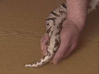
The cloaca can be examined very simply by using a otoscope or endoscope. Check for oedema, erythema, discharge and swellings. Check the vent for any soiling which may be due to parasitic infection of the large intestine. Make a faecal smear if possible. Examine the tail for retained sloughed skin, lumps and other deformities.
Sexing/Probing
The snake is positioned so that the vent is easily accessible. A smooth lubricated blunt-tipped probe is inserted caudally through the vent. In males the probes enters the hemipenes (10-12 scale rows) while in females it may enter the musk glands (2-3 scale rows). Take care since rough use of probes can cause damage. In females it is possible to perforate the musk gland and allow the probe to slide in as far as it would in a male snake.
References
Fowler, M.E. and Miller, R.E. (2003). Zoo and Wild Animal Medicine. Saunders, 5th Edition. pp. 84. ISBN 0-7216-9499-3
