Difference between revisions of "Snake Respiratory System"
| Line 1: | Line 1: | ||
{{review}} | {{review}} | ||
| − | [[Image:718099 - external_nares_ed_copy.jpg|200px|thumb|right|'''External nares''' | + | [[Image:718099 - external_nares_ed_copy.jpg|200px|thumb|right|'''External nares''' © RVC]] |
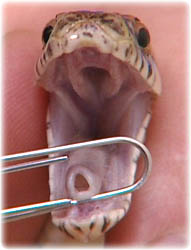
The respiratory tract of snakes consists of external nares, nasal cavity, internal nares, glottis, trachea, bronchi, lung(s) and air sac. There is no diaphragm. The external nares communicate with the internal nares through the nasal cavity. The internal nares lie in the roof of the oral cavity and communicate directly with the glottis when the mouth is closed. The glottis lies posterior to the tongue and is not difficult to visualise, making intubation for [[Lizard and Snake Anaesthesia|anaesthesia]] relatively easy. During the feeding process, the glottis is able to move laterally to facilitate respiration while ingesting large prey items, a process which may take considerable time. | The respiratory tract of snakes consists of external nares, nasal cavity, internal nares, glottis, trachea, bronchi, lung(s) and air sac. There is no diaphragm. The external nares communicate with the internal nares through the nasal cavity. The internal nares lie in the roof of the oral cavity and communicate directly with the glottis when the mouth is closed. The glottis lies posterior to the tongue and is not difficult to visualise, making intubation for [[Lizard and Snake Anaesthesia|anaesthesia]] relatively easy. During the feeding process, the glottis is able to move laterally to facilitate respiration while ingesting large prey items, a process which may take considerable time. | ||
| − | + | '''For more information, see''' [[Snake Special Senses]]. | |
| − | + | '''For more information on physical examinations, see''' [[Snake Physical Examination]]. | |
==Trachea== | ==Trachea== | ||
| − | [[Image:Glottis_&_int_nares_copy.jpg|200px|thumb|right|'''Internal nares and glottis''' | + | [[Image:Glottis_&_int_nares_copy.jpg|200px|thumb|right|'''Internal nares and glottis''' © RVC]] |
The trachea has incomplete cartilaginous rings. The ventral portion is rigid cartilage while the dorsal fourth is membranous. The trachea enters the lung at a level near the base of the heart. Some species of snakes have a unique saccular diverticulum of open tracheal rings that acts for hissing or as a tracheal lung for gas exchange when gastric contents preclude normal pulmonary function. | The trachea has incomplete cartilaginous rings. The ventral portion is rigid cartilage while the dorsal fourth is membranous. The trachea enters the lung at a level near the base of the heart. Some species of snakes have a unique saccular diverticulum of open tracheal rings that acts for hissing or as a tracheal lung for gas exchange when gastric contents preclude normal pulmonary function. | ||
==Lungs== | ==Lungs== | ||
The lung morphology of snakes is very species-dependent but is obviously elongated. The left lung is always reduced but may vary from 85% of the length of the right lung to being absent. It is relatively well developed in [[Boidae|boids]] but only vestigial in [[Colubridae|colubrids]]. The anterior part of the lung is non-respiratory and functions as an air sac for gas exchange. In [[Boidae|boids]] and [[Colubridae|colubrids]], the respiratory portion of the lung lies between the [[Snake Cardiovascular System|heart]] and the cranial liver whereas in most [[Viperidae|viperids]] and [[Elapidae|elapids]] it is cranial to the [[Snake Cardiovascular System|heart]]. The posterior part is thin-walled and non-respiratory, mainly functions as an air sac and contains a major portion of the volume of the respiratory tract. In terrestrial snakes this air sac terminates in the intestinal mesentery near the gallbladder while in aquatic species it may reach to the [[Cloaca|cloaca]] and is thicker and more muscular. | The lung morphology of snakes is very species-dependent but is obviously elongated. The left lung is always reduced but may vary from 85% of the length of the right lung to being absent. It is relatively well developed in [[Boidae|boids]] but only vestigial in [[Colubridae|colubrids]]. The anterior part of the lung is non-respiratory and functions as an air sac for gas exchange. In [[Boidae|boids]] and [[Colubridae|colubrids]], the respiratory portion of the lung lies between the [[Snake Cardiovascular System|heart]] and the cranial liver whereas in most [[Viperidae|viperids]] and [[Elapidae|elapids]] it is cranial to the [[Snake Cardiovascular System|heart]]. The posterior part is thin-walled and non-respiratory, mainly functions as an air sac and contains a major portion of the volume of the respiratory tract. In terrestrial snakes this air sac terminates in the intestinal mesentery near the gallbladder while in aquatic species it may reach to the [[Cloaca|cloaca]] and is thicker and more muscular. | ||
| − | + | '''For more information, see''' [[Lizard and Snake Respiration]] '''and''' [[Snake Respiratory System]]. | |
| − | |||
| − | |||
[[Category:Snake_Anatomy]] | [[Category:Snake_Anatomy]] | ||
Revision as of 14:45, 18 May 2010
| This article has been peer reviewed but is awaiting expert review. If you would like to help with this, please see more information about expert reviewing. |
The respiratory tract of snakes consists of external nares, nasal cavity, internal nares, glottis, trachea, bronchi, lung(s) and air sac. There is no diaphragm. The external nares communicate with the internal nares through the nasal cavity. The internal nares lie in the roof of the oral cavity and communicate directly with the glottis when the mouth is closed. The glottis lies posterior to the tongue and is not difficult to visualise, making intubation for anaesthesia relatively easy. During the feeding process, the glottis is able to move laterally to facilitate respiration while ingesting large prey items, a process which may take considerable time.
For more information, see Snake Special Senses.
For more information on physical examinations, see Snake Physical Examination.
Trachea
The trachea has incomplete cartilaginous rings. The ventral portion is rigid cartilage while the dorsal fourth is membranous. The trachea enters the lung at a level near the base of the heart. Some species of snakes have a unique saccular diverticulum of open tracheal rings that acts for hissing or as a tracheal lung for gas exchange when gastric contents preclude normal pulmonary function.
Lungs
The lung morphology of snakes is very species-dependent but is obviously elongated. The left lung is always reduced but may vary from 85% of the length of the right lung to being absent. It is relatively well developed in boids but only vestigial in colubrids. The anterior part of the lung is non-respiratory and functions as an air sac for gas exchange. In boids and colubrids, the respiratory portion of the lung lies between the heart and the cranial liver whereas in most viperids and elapids it is cranial to the heart. The posterior part is thin-walled and non-respiratory, mainly functions as an air sac and contains a major portion of the volume of the respiratory tract. In terrestrial snakes this air sac terminates in the intestinal mesentery near the gallbladder while in aquatic species it may reach to the cloaca and is thicker and more muscular.
For more information, see Lizard and Snake Respiration and Snake Respiratory System.

