Spinal Column - Anatomy & Physiology
Jump to navigation
Jump to search
BACK TO MUSCULOSKELETAL ANATOMY AND PHYSIOLOGY
BACK TO MUSCULOSKELETAL ANATOMY AND PHYSIOLOGY
Divisions and Landmarks
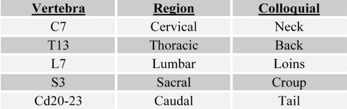
The common pattern of canine landmarks, according to Dyce, Sack, et al, can be quantified as such:


Vertebrae and Joints
- Vertebrae consist of a body which encloses the vertebral foramen (through which the spinal cord and meninges run), a spinous process, and a transverse process, as well as articular processes by which they join together
- The form of the spinous process varies with respect to species and region
Cervical Vertebrae
- The first two cervical vertebrae are known as the atlas and the axis respectively, and are modified to allow movement of the head
- The atlas has no conventional body: instead it is complsed of two lateral masses joined by dorsal and ventral arches
- The atlas and axis are fused in embryonic life
- The wing of the atlas is the transverse process of this vertebra and allows the spinal column to articulate with the skull by providing a resting place for the occipital condyles
- The axis is the longest vertebra
- The nuchal ligament connects the spinous process of the axis to the spinous process of the first thoracic vertebra (T1)
- The last (C7) cervical vertebra has a taller spinous process than those proceeding it, and articulates with the first pair of ribs
Thoracic Vertebrae
- Thoracic vertebrae articulate with the ribs
- They are distingushed by short bodies with flattened extremities, costal facets, short transverse processes, and prominent spinous processes
- They reach a maximum height a few vertebrae behind the cervicothoracic junction (constituting the withers of the horse) and then decline
- The orientation of spinous processes shifts from caudo- to craniodorsal
Lumbar Vertebrae
- Longer and more uniform in shape than thoracic vertebrae
- Shorter in height, with long, flattened transverse processes that project laterally
Sacral Vertebrae
- Sacrum: a single bone formed by the fusion of several vertebrae that articulates with the pelvic girdle
- Allows the thrust of the hindlimbs to be transmitted to the trunk
- Narrows caudally and is curved to present a concave surface to the pelvic cavity
Caudal Vertebrae
- Number varies greatly even within species
- Progressive simplification of form
Joints of the Spinal Column
- Two types of joints:
- Cartilaginous: direct connections between vertebral bodies
- Bodies of adjacent vertebrae connected by thick, flexible intervertebral discs, consisting of two parts:
- Nucleus pulposus: slightly eccentric, notochord derivative, contained under pressure and prone to escape
- Annulus fibrosis: encircling bundles of fibrous tissue that pass obliquely from one vertebra to another, with changing orientation
- Bodies of adjacent vertebrae connected by thick, flexible intervertebral discs, consisting of two parts:
- Synovial: between facets on vertebral arches
- Cartilaginous: direct connections between vertebral bodies
- Modified in the regions of the head and pelvis
- Joints of the atlas:
- Atlanto-occipital joint: between condyles of the skull and corresponding cavities of the atlas
- Functions as a ginglymus: movement is restricted to flexion/extension in the sagittal plane (eg nodding)
- Atlantoaxial joint: ventral arch of atlas and the body of the axis face into a single synovial cavity with limited areas of contact
- Movement is rotational about a longitudinal axis (eg. head shaking)
- Atlanto-occipital joint: between condyles of the skull and corresponding cavities of the atlas
- Joints of the atlas:
Spinal Cord
Hypaxial and Epaxial Muscles

- Epaxial muscles: extensors of the vertebral column
- Dorsal to the line of the transverse processes of the vertebrae
- Innervation comes from the dorsal branches of the spinal nerves
- Rarely of clinical importance
- Arranged in three parallel columns
- Lateral column: Iliocostalis arises from the ilium and transverse processes of the lumbar vertebrae to insert on cranial lumbar vertebrae and ribs, spanning about 4 vertebrae
- Middle column: Longissimus; strongest, extending from the ilium and sacrum to the head and neck
- Medial column: Transversospinalis; most complex, lying between medial vertebral arches and spinous processes

- Hypaxial Muscles: flexors of the neck and tail
- Longus colli: from cranial thoracic region to the atlas, covering ventral vertebral bodies
- Rectus capitis ventralis: atlas to ventral skull
- Longus capitis: midcervical vertebrae to skull
- Scalenus muscles: caudal cervica vertebrae to first few ribs, which they stabilize on inspiration