Difference between revisions of "Stomatitis"
| Line 1: | Line 1: | ||
| + | {{unfinished}} | ||
{{cat}} | {{cat}} | ||
{{dog}} | {{dog}} | ||
| Line 94: | Line 95: | ||
**Result of other more systemic diseases | **Result of other more systemic diseases | ||
[[Category:Oral_Cavity_and_Gingiva_-_Pathology]] | [[Category:Oral_Cavity_and_Gingiva_-_Pathology]] | ||
| − | [[Category:To_Do_- | + | [[Category:To_Do_-_Caz]] |
Revision as of 20:50, 26 July 2010
| This article is still under construction. |
Typical Signalment
- Both dogs and cats can develop this condition
- Breeds of dog predisposed include:
 Greyhound
ToB 2005, WikiMedia Commons
 Maltese
Sannse 2003, WikiMedia Commons
 Cavalier King Charles Spaniel
Andreweatock 2009, WikiMedia Commons
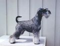 Miniature Schnauzer
MagnusK 2006, WikiMedia Commons
 Retriever (Labrador)
Elf 2004, WikiMedia Commons


Description
Stomatitis is the inflammation of the mucosa lining any structures within the mouth. This may include the lips, cheeks, tongue and gingiva.
Several different types of stomatitis have been described in the dog:
- Chronic ulcerative paradental stomatitis - Seen on the buccal mucosa that overlie the teeth, especially in the area of the maxillary canine tooth, fourth premolar and the lateral edge of the tongue. Severe ulceration can occur together with gingival recession.
- Ulcerative stomatitis - Ulcerations on the margins of the tongue are common together with a secondary lip-fold dermatitis due to excessive salivation.
- Necrotizing stomatitis - A very painful condition in dogs that may be caused by opportunistic invasion of normal oral flora. Suspected causative organisms include Fusobacterium and spirochaetes. Invasion of these organisms is thought to be associated with reduced host resistance.
- Uraemic stomatitis - Occurs as a result of uraemia due to renal disease. Severe stomatitis and ulceration of the oral mucosa as well as the margins of the tongue are seen with this condition. The lesions occur due to the bacterial degradation of urea to form ammonia together with dehydration and drying of the oral mucosa that results from renal disease.
Stomatitis in the cat:
Diagnosis
Clinical Signs
- severe halitosis
- hypersalivation
- thick, ropey saliva
- anorexia caused by pain
- malaise
- febrile
- weight loss
- ulceration and bleeding of the gingiva.
Oral Examination
A thorough oral examination under general anaesthesia is often required to aid the diagnosis. Diagnosis is usually made by gross visualisation of the lesions. The mandibular lymph nodes may also be enlarged.
Laboratory Tests
Clinical pathology may be carried out to assess renal function in the case of uraemic stomatitis. Culture and sensitivity tests may be required to aid diagnosis and enable specific treatment.
Biopsy
Often indicated to rule out other diseases such as pemphigus or other immunopathies.
Treatment
Treatment is both symptomatic to control the clinical signs as well as specific to the cause of the condition. The underlying cause should be sought and treated appropriately.
- Thorough teeth cleaning
- Systemic antibiotics (effective against aeobes and anaerobes)
- Oral rinses such as chlorhexidine
- Corticosteriods have been suggested with cases of chronic ulcerative paradental stomatitis.
- Diet modification in cases of renal disease to reduce blood urea levels.
- Tooth extraction in the most severely affected area has been suggested.
- Resistent lesions in cats have been shown to respond to Bovine lactoferrin in some cases.
Prognosis
Depends on the underlying cause.
References
- Merck & Co (2008) The Merck Veterinary Manual
- Nelson, R.W. and Couto, C.G. (2009) Small Animal Internal Medicine (Fourth Edition) Mosby Elsevier
- Tutt, C., Deeprose, J. and Crossley, D. (2007) BSAVA Manual of Canine and Feline Dentistry (3rd Edition) BSAVA
From Pathology
Catarrhal Stomatitis
- Non-specific, general stomatitis
Pathology
- Starts as hyperaemia and oedema of tongue or pharynx with mucoid exudate on surface.
- Lymphoid follicles on soft palate may enlarge and proliferate.
- Often see white spots due to epithelial hyperplasia and increased mucous secretion.
- (can be scraped off to leave ordinary mucosa underneath).
- May produce bad smell.
- Resolves normally if not secondarily infected.
Pathogenesis
- May be caused by:
- Low grade streptococcal infection
- Ingestion of toxins
- Result of other more systemic diseases