Thymus - Anatomy & Physiology

Introduction
The thymus has a key role in the maturation of prothymocytes into mature T cells. In juvenile animals the thymus produces significant numbers of new T lymphocytes but as the animal matures this production decreases and T cell population is maintained by division of mature T cells.
Embryology
Note: Embryology here is brief and simplified
The thymus mainly develops from the left and right third pharyngeal pouches and extends caudally until it fuses with the pericardium. Then as the heart migrates into the thoracic cavity the thymus comes with it. At this point the thymus is Y-shaped and this shape persists in neonatal ruminants, in horses the left-right connection is lost leaving two separate lobes while in carnivores only the thoracic part remains and the cranial portion regresses.
During development some cells (CFU-L’s) migrate from the bone marrow to the thymus and eventually become concentrated around the outside of the thymus, producing a dense layer of cells (the cortex) and giving rise to the inner medulla. Some of the cells in the medulla surround and form layers around enlarged endodermal cells and these give rise to the Hassall’s corpuscles.
Structure
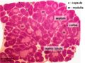
The thymus is found cranial to the heart and has a 'lobular' strucutre. Within the lobules the tissue consists of an outer cortex and an inner medulla.
General structure
The thymus is surrounded by a capsule made of thin connective tissue. Trabeculae from this extend into the thymus creating thymic ‘lobules’. The connective tissues house blood vessels and efferent lymphatic vessels along with nerves.
The cortex is dense consisting of rapidly dividing thymocytes (developing T lymphocytes) which stain with H&E to give a basophilic appearance. The thymocytes are supported in a network of epithelioreticular cells (‘epithelio’ due to similarities with epithelium and ‘reticular’ due to similarity with reticular cells in lymph tissues). Small numbers of dendritic epithelial cells are also present.
There are six types of epithelioreticular cells. Types I-III are found in the cortex and types IV-VI in the medulla. Type VI forming Hassall’s (or thymic) corpuscles.
The medulla has non-dividing, more mature T cells with dendritic cells at the cortico-medullary junction. The density of thymocytes is much less than the cortex and consequently it does not stain as strongly. The cytoplasm in Hassall’s corpuscles contains keratohyalin granules and intermediate fibres and may be keratinised. Function is yet to be asserted but thought to produce IL-4 and IL-7 to help with T lymphocyte development.
Blood-thymus barrier
The blood-thymus barrier protects the T lymphocytes from exposure to antigens in the blood. It is formed in the capillaries by a continuous endothelium with occluding junctions surrounded by connective tissue and then surrounded by a second layer formed from the processes of epithelioreticular cells (Type I).
Macrophages are also present around the capillaries to engulf any antigenic substances that manage to penetrate the barrier.
Changes
The thymus remains a sizeable organ until the animal reaches puberty. Once puberty is reached the lymphoid tissue is replaced with adipose tissue (involution).
Histology
Low power magnification
©RVC 2008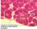
Medulla, Cotex & Capsule
©RVC 2008
Medulla & Cortex
©RVC 2008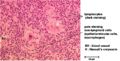
Hassall’s corpuscle
©RVC 2008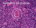
Hassall’s corpuscle
©Nottingham Uni 2008


Functions
The thymus has a key role in the maturation of prothymocytes into mature T cells. In juvenile animals the thymus produces significant numbers of new T lymphocytes, but as the animal matures this production decreases and the T cell population is maintained by division of mature T cells.
Endocrine
Although the Thymus shrinks after puberty, it continues to function as an endocrine gland during adulthood. It produces the hormone thymosin which stimulates the activity of the T lymphocytes.
Back to Endocrine System
Links
| Thymus - Anatomy & Physiology Learning Resources | |
|---|---|
Anatomy Museum Resources |
Canine Thymus Histology Avian Thymus Histology |
| Originally funded by the RVC Jim Bee Award 2007 |
Webinars
Failed to load RSS feed from https://www.thewebinarvet.com/internal-medicine/webinars/feed: Error parsing XML for RSS