Difference between revisions of "Trachea - Anatomy & Physiology"
Ggaitskell (talk | contribs) |
|||
| (39 intermediate revisions by 9 users not shown) | |||
| Line 1: | Line 1: | ||
| − | {{ | + | {{OpenPagesTop}} |
| − | |||
| − | |||
| − | |||
| − | |||
| − | |||
| − | }} | ||
| − | |||
| − | |||
| − | |||
| − | |||
| − | |||
==Introduction== | ==Introduction== | ||
| − | The trachea is the tube linking the cricoid [[ | + | The trachea is the tube linking the cricoid [[Cartilage - Anatomy & Physiology#Types of Cartilage|cartilage]] of the [[Larynx - Anatomy & Physiology |larynx]] to the [[Bronchi and Bronchioles - Anatomy & Physiology|bronchi]], forming part of the conducting system which transports air from the external environment to the [[Lungs - Anatomy & Physiology|lungs]]. The [[Oesophagus - Anatomy & Physiology|oesophagus]] lies dorsally to the trachea. The cervical part of the trachea lies generally in the median position, although this varies slightly depending on the position of the head. The thoracic part of the trachea crosses the aortic arch, thus its positioning is moved slightly to the right at this level. The trachea bifurcates to form the two [[Bronchi and Bronchioles - Anatomy & Physiology|bronchi]] at the level of the 4th-6th intercostal space. |
| − | The cervical part of the trachea lies generally in the median position, although this varies slightly depending on the position of the head. | ||
| − | The thoracic part of the trachea crosses the aortic arch, thus | ||
| − | |||
| − | The trachea bifurcates to form the two [[Bronchi and | ||
==Structure== | ==Structure== | ||
| − | + | The trachea contains numerous rings of [[Cartilage - Anatomy & Physiology#Hyaline Cartilage|hyaline cartilage]] which are C-shaped, being dorsally incomplete, connected to each other by elastic connective tissue. The ends of the incomplete rings are joined by the smooth ''trachealis'' muscle. The structural conformation of the trachea prevents collapse due to traction forces, whilst allowing it to adjust in length and diameter, as the neck moves and the [[Diaphragm - Anatomy & Physiology|diaphragm]] contracts. The trachea's walls are made up of a number of layers including the inner mucosa, fibrocartilaginous middle layer, and adventitia (in the neck) or serosa (in the thorax). The inner mucosa contains glands which produce mucus. This mucus traps debris and is constantly moved upwards towards the [[Oropharynx - Anatomy & Physiology|oropharynx]] where it is swallowed. This mechanism is known as the [[Respiratory Epithelium - Anatomy & Physiology#Mucociliary Escalator|'''Muco-Ciliary escalator''']]. | |
| − | |||
| − | |||
| − | |||
==Function== | ==Function== | ||
| − | + | The trachea is responsible for transporting air for respiration from the [[Larynx - Anatomy & Physiology|larynx]] to the [[Bronchi and Bronchioles - Anatomy & Physiology|bronchi]]. | |
| + | |||
| + | ==Species Differences== | ||
| + | |||
| + | In the '''dog and cat''' the C-Shaped rings are joined by muscle which is placed externally, rather than internally as is normal for the other species. In [[Avian Respiration - Anatomy & Physiology|'''avian]] species''' the trachea is composed of tightly stacked rings of [[Cartilage - Anatomy & Physiology#Types of Cartilage|cartilage]], which are complete with no dorsal space. They overlap considerably. The [[Respiration in Non-Homeotherms - Anatomy & Physiology|respiratory systems of '''non-homeotherms''']] are also very different to that of mammals. | ||
==Histology== | ==Histology== | ||
<center><gallery> | <center><gallery> | ||
| − | Image: | + | Image:TracheaRatlowpower.jpg |<p>A histology section of a trachea (rat) <sub>©RVC 2008</sub></p> |
| − | Image:TrachealWallLiningRAT.jpg|<p> | + | Image:TrachealWallRAT.jpg|<p>A histology section of the tracheal wall (rat) <sub>©RVC 2008</sub></p> |
| − | Image:TrachealWallstainedforelasticfibresCAT.jpg|<p> | + | Image:TrachealWallLiningRAT.jpg|<p>A histology section of the epithelial lining of the tracheal wall (rat) <sub>©RVC 2008</sub></P> |
| + | Image:TrachealWallstainedforelasticfibresCAT.jpg|<p>A histology section of the tracheal wall stained for elastic fibres (Cat)</P> <sub>©RVC 2008</sub>]] | ||
</gallery></center> | </gallery></center> | ||
| − | |||
| − | |||
| − | |||
| − | |||
| − | |||
==Links== | ==Links== | ||
| + | Click here for information on [[:Category:Trachea - Pathology|trachea pathology]]. | ||
| + | <br> | ||
| + | {{Learning | ||
| + | |flashcards= [[Syrinx - Anatomy & Physiology|The syrinx and avian vocalisation]] | ||
| + | |powerpoints= [[Respiratory System Histology resource|Histology of the respiratory system, including the trachea]] | ||
| + | |dragster = [[Respiratory System Histology Resource (I)|Trachea Histology Dragster]] | ||
| + | |OVAM = [http://www.onlineveterinaryanatomy.net/content/respiration-histology-images-trachea-1 Histology - Trachea 1]<br>[http://www.onlineveterinaryanatomy.net/content/respiration-histology-images-trachea-2 Histology - Trachea 2]<br>[http://www.onlineveterinaryanatomy.net/content/respiration-histology-images-trachea Histology - Trachea 3]<br>[http://www.onlineveterinaryanatomy.net/content/respiration-histology-images-trachea-0 Histology - Trachea 4]<br>[http://www.onlineveterinaryanatomy.net/content/equine-trachea-histology Histology - Equine Trachea]<br>[http://www.onlineveterinaryanatomy.net/content/canine-trachea-histology Histology - Canine Trachea] | ||
| + | }} | ||
| + | <br> | ||
| − | + | ==References== | |
| − | == | + | {{citation|initiallast = Dyce|initialfirst = K.M|2last = Sack|2first = W.O|finallast = Wensing|finalfirst = C.J.G|year = 2002|title = Textbook of Veterinary Anatomy|ed =3rd|city = Philadelphia|pub = Saunders}} |
| − | + | {{review}} | |
| + | {{OpenPages}} | ||
| + | [[Category:Respiratory System - Anatomy & Physiology]] | ||
| + | [[Category:A&P Done]] | ||
Revision as of 06:38, 21 August 2014
Introduction
The trachea is the tube linking the cricoid cartilage of the larynx to the bronchi, forming part of the conducting system which transports air from the external environment to the lungs. The oesophagus lies dorsally to the trachea. The cervical part of the trachea lies generally in the median position, although this varies slightly depending on the position of the head. The thoracic part of the trachea crosses the aortic arch, thus its positioning is moved slightly to the right at this level. The trachea bifurcates to form the two bronchi at the level of the 4th-6th intercostal space.
Structure
The trachea contains numerous rings of hyaline cartilage which are C-shaped, being dorsally incomplete, connected to each other by elastic connective tissue. The ends of the incomplete rings are joined by the smooth trachealis muscle. The structural conformation of the trachea prevents collapse due to traction forces, whilst allowing it to adjust in length and diameter, as the neck moves and the diaphragm contracts. The trachea's walls are made up of a number of layers including the inner mucosa, fibrocartilaginous middle layer, and adventitia (in the neck) or serosa (in the thorax). The inner mucosa contains glands which produce mucus. This mucus traps debris and is constantly moved upwards towards the oropharynx where it is swallowed. This mechanism is known as the Muco-Ciliary escalator.
Function
The trachea is responsible for transporting air for respiration from the larynx to the bronchi.
Species Differences
In the dog and cat the C-Shaped rings are joined by muscle which is placed externally, rather than internally as is normal for the other species. In avian species the trachea is composed of tightly stacked rings of cartilage, which are complete with no dorsal space. They overlap considerably. The respiratory systems of non-homeotherms are also very different to that of mammals.
Histology
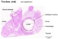
A histology section of a trachea (rat) ©RVC 2008
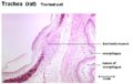
A histology section of the tracheal wall (rat) ©RVC 2008
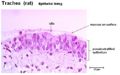
A histology section of the epithelial lining of the tracheal wall (rat) ©RVC 2008
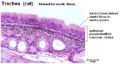
A histology section of the tracheal wall stained for elastic fibres (Cat)
©RVC 2008]]


Links
Click here for information on trachea pathology.
| Trachea - Anatomy & Physiology Learning Resources | |
|---|---|
Test your knowledge using drag and drop boxes |
Trachea Histology Dragster |
Test your knowledge using flashcard type questions |
The syrinx and avian vocalisation |
 Selection of relevant PowerPoint tutorials |
Histology of the respiratory system, including the trachea |
Anatomy Museum Resources |
Histology - Trachea 1 Histology - Trachea 2 Histology - Trachea 3 Histology - Trachea 4 Histology - Equine Trachea Histology - Canine Trachea |
References
Dyce, K.M., Sack, W.O. and Wensing, C.J.G. (2002) Textbook of Veterinary Anatomy. 3rd ed. Philadelphia: Saunders.
| This article has been peer reviewed but is awaiting expert review. If you would like to help with this, please see more information about expert reviewing. |
Error in widget FBRecommend: unable to write file /var/www/wikivet.net/extensions/Widgets/compiled_templates/wrt661f08b86ca6d2_07000237 Error in widget google+: unable to write file /var/www/wikivet.net/extensions/Widgets/compiled_templates/wrt661f08b871dbc8_52870445 Error in widget TwitterTweet: unable to write file /var/www/wikivet.net/extensions/Widgets/compiled_templates/wrt661f08b87e04f3_77521852
|
| WikiVet® Introduction - Help WikiVet - Report a Problem |