|
Also Known As: Canker — Frounce — Roup — Tetratrichomiasis — Trichomoniasis — Avian Trichomonosis
Caused by: Trichomonas gallinae
Introduction
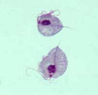
Avian trichomonosis is primarily an upper gastrointestinal disease of birds caused by the flagellate protozoal pathogen Trichomonas gallinae.
Columbiformes (pigeons and doves) are the primary hosts. Residing primarily in the crop or ingluvies, the parasite is passed from infected parents to offspring during “pigeon milk” feeding early in life. Adult columbiformes also transmit the protozoan during courtship displays. Infection can then persist for considerable time. Only young or debilitated birds usually show signs of disease.
Domestic chicken and turkey infections are often acquired from drinking water contaminated by wild birds, usually feral pigeons, Columba livia or Wood Pigeons, Columba palumbus. There is no vertical transmission from parent to offspring. Birds of prey can also acquire the disease by ingestion of infected birds, and in these species, the disease is often referred to as frounce.
Distribution
Trichomonosis is present worldwide and is a significant cause of economic loss in pigeons and turkeys. T. gallinae is an exceptionally hardy parasite, and can survive a wide range of suboptimal environmental conditions without a cyst form.
Clinical Signs
Presence of the parasite in the crop, oesophagus, mouth, throat and sinuses causes yellow lesions/masses that grow in size and become obstructive. Birds may not be able to close their mouths, causing dyspnoea, dysphagia and fluid accumulation within the oral cavity. Weight loss, ocular discharge, ataxia and blindness are also commonly reported.
Diagnosis
Presence of yellow vesicles, “buttons” or masses on the oral mucosa and oesophagus is highly suggestive.
Pharyngeal or crop swabs can be used for laboratory culture and Giemsa staining achieving definitive diagnosis. Immediate microscopy of a direct swab may reveal motile, flagellated protozoal organisms.
Pathology
Caseous lesions/masses are commonly present within the mouth, oesophagus, crop and glandular stomach.
The most frequently affected visceral organ is the liver, which can then occasionally cause distribution throughout the abdominal cavity.
In very young pigeons (squabs) the navel region may be affected, encasing many visceral organs in a caseous mass.
Treatment
2-amino-5-nitrothiazole is the drug of choice due to its lack of adverse effects and few resistance problems at present.
Carnidazole, dimetridazole, metronizadole and ronidazole have also been used successfully, although attention should be paid when administering these drugs to food producing animals. All are administered orally, either in liquid or tablet form for direct medication, or dissolved in water.
Control
Prevention of wild birds access to water sources, good sanitation protocols and quarantine of new birds are paramount to control of this disease.
Relevant links: Bovine Trichomonosis
| Trichomonosis - Birds Learning Resources | |
|---|---|
 Test your knowledge using flashcard type questions |
Avian Trichomonosis Flashcards |
 Search for recent publications via CAB Abstract (CABI log in required) |
Avian Trichomonosis Publications |
References

|
This article was originally sourced from The Animal Health & Production Compendium (AHPC) published online by CABI during the OVAL Project. The datasheet was accessed on 3 June 2011. |
BonDurant, R.H., Honigberg, B.M., 1994. Trichomonads of veterinary importance. In: Kreier, J.P. (Ed.), Parasitic Protozoa, 2nd ed. vol. 9. Academic Press, San Diego, CA, 168–177.
Frank K. (2004). Canker [online] Available at: http://www.albertaclassic.com/trichomonas/trichomonas.php. [Accessed 12 May 2011]
Franssen, F. F. J., Lumeij, J. T.(1992) In vitro nitroimidazole resistance of Trichomonas gallinae and successful therapy with an increased dosage of ronidazole in racing pigeons (Columba livia domestica). J Vet Pharm Therap, 15(4):409-415; 20.
Levine N. D (1973) The Trichomonads Levine ND, ed. Protozoan Parasites of Domestic Animals and of Man. Burgess Publishing Company. Minnesota, USA , 88-110.
Samour J. H., Bailey, T. A., Cooper, J. E. (1995) Trichomoniasis in birds of prey (Order falconiformes) in Bahrain. Vet Record, 136(14):358-362.
| This article has been expert reviewed by Dr Tiana Tasca, Dr De Carli & Dr. Bev Panto. Date reviewed: 27th July 2017 |
Error in widget FBRecommend: unable to write file /var/www/wikivet.net/extensions/Widgets/compiled_templates/wrt6620b21e7d4921_02908304 Error in widget google+: unable to write file /var/www/wikivet.net/extensions/Widgets/compiled_templates/wrt6620b21e8532c1_93030979 Error in widget TwitterTweet: unable to write file /var/www/wikivet.net/extensions/Widgets/compiled_templates/wrt6620b21e8944a5_96497179
|
| WikiVet® Introduction - Help WikiVet - Report a Problem |

