Difference between revisions of "Urinary Bladder - Anatomy & Physiology"
| (48 intermediate revisions by 6 users not shown) | |||
| Line 1: | Line 1: | ||
| − | {{ | + | {{OpenPagesTop}} |
| − | + | ==Introduction== | |
| − | + | * The bladder is where urine is stored before being expelled by the body through the [[Micturition - Anatomy & Physiology| micturition reflex]]. | |
| − | |||
| − | |||
| − | |||
| − | |||
| − | |||
| − | |||
| − | |||
| − | * The bladder is where urine is stored before being expelled by the body through the [[ | ||
* Without a bladder urinary continence would be impossible. | * Without a bladder urinary continence would be impossible. | ||
==Anatomy== | ==Anatomy== | ||
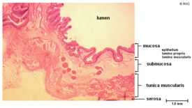
| − | + | [[Image:bladhistoanat1.jpg|right|thumb|275px|<small><center>Histology cross section of a normal guinea pig bladder (© RVC 2008)</center></small>]] | |
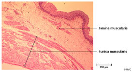
| + | [[Image:bladhistoanat2.jpg|right|thumb|275px|<small><center>Histology cross section of a normal guinea pig bladder (© RVC 2008)</center></small>]] | ||
* It is a hollow, muscular organ | * It is a hollow, muscular organ | ||
* It is divided for descriptive purposes into three parts | * It is divided for descriptive purposes into three parts | ||
| Line 19: | Line 12: | ||
** Intermediate body | ** Intermediate body | ||
** Caudal neck | ** Caudal neck | ||
| − | * Its wall comprises a muscle layer covered in | + | * Its wall comprises a muscle layer covered in transitional epithelium. |
* Its size and posistion are determined by how full it is. | * Its size and posistion are determined by how full it is. | ||
** When empty the bladder wall is wrinkled and thicker | ** When empty the bladder wall is wrinkled and thicker | ||
| − | ** When full and distended the folds | + | *** It rests on the pubic bones |
| − | * The paired uteric folds | + | *** It is entirely in the pelvis of the large species but partly enters the abdomen of the carnivores |
| − | ** | + | *** It is largely retroperitoneal in the larger species |
| − | ** | + | ** When full and distended the folds disappear and the wall appears thinner |
| − | ** | + | *** It then becomes intraperitoneal in the larger species |
| + | * The '''trigone''' of the bladder gets its name as it looks like a triangle without a base. | ||
| + | ** It is of clinical importance and is formed by the paired uteric folds | ||
| + | ** These form by the orifice of the ureters | ||
| + | ** It is visible even when the bladder is full | ||
| + | ** The folds extend from the urethral opening to the neck of the bladder | ||
| + | ** Where they merge to form the urethral crest | ||
| + | ** It is believed to have increased sensitivty and is of differant embryological origin to the rest of tissue. More details can be found [[Kidney and Urinary Tract Development - Anatomy & Physiology#The Bladder|here]] | ||
| − | + | ==Muscles of the Bladder== | |
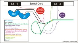
| − | The three muscular components of the bladder described below play a pivotal part in the [[ | + | [[Image:sumlutshcemtri.jpg|right|thumb|275px|<small><center>A schematic overview of the lower urinary tract showing the innervation and muscles of the bladder</center></small>]] |
| + | The three muscular components of the bladder described below play a pivotal part in the [[Micturition - Anatomy & Physiology| micturition reflex.]] | ||
====Detrusor Muscle==== | ====Detrusor Muscle==== | ||
| − | + | This network of smooth muscle fibres lie in three sheets within the bladder wall and are supplied by both parasympathetic and sympathetic nerves. It is responsible for storage and expression of urine from the bladder. | |
=====Parasympathetic Supply - Detrusor Muscle===== | =====Parasympathetic Supply - Detrusor Muscle===== | ||
* S1-S3 | * S1-S3 | ||
* Synapse in pelvic plexus or bladder wall | * Synapse in pelvic plexus or bladder wall | ||
| + | * Pelvic nerves | ||
* Innvervate the detrusor muscle | * Innvervate the detrusor muscle | ||
* Action - excitatory | * Action - excitatory | ||
* Function - empty bladder | * Function - empty bladder | ||
| + | |||
=====Sympathetic Supply - Detrusor Muscle===== | =====Sympathetic Supply - Detrusor Muscle===== | ||
* L1-L4 | * L1-L4 | ||
* Syanpse in caudal mesenteric ganglion - bladder wall | * Syanpse in caudal mesenteric ganglion - bladder wall | ||
| − | * Receptor - beta | + | * Hypogastric nerve |
| + | * Receptor - beta 2 | ||
* Inhibitory action | * Inhibitory action | ||
* Allows bladder filling | * Allows bladder filling | ||
====Internal Urethral Sphincter==== | ====Internal Urethral Sphincter==== | ||
| − | + | A thickening of the bladder musculature found at the neck of the bladder which is continuous with the detrusor and is therefore smooth muscle. However, unlike the detrusor its innervation is purely from sympathetic fibres. | |
=====Sympathetic Supply - Internal Urethral Sphincter===== | =====Sympathetic Supply - Internal Urethral Sphincter===== | ||
* L1-L4 | * L1-L4 | ||
* Synapse in caudal mesenteric ganglion | * Synapse in caudal mesenteric ganglion | ||
| − | * Receptor - alpha | + | * Hypogastric nerve |
| + | * Receptor - alpha 1 | ||
* Excitatory action | * Excitatory action | ||
* Function - retain urine through increased urethral tone | * Function - retain urine through increased urethral tone | ||
====External Urethral Sphincter==== | ====External Urethral Sphincter==== | ||
| − | + | This third component is more appropriately included in the anatomy section of the [[Urethra - Anatomy & Physiology#Muscles of the Urethra|urethra]]. | |
| − | |||
| − | |||
| − | |||
| − | |||
| − | |||
| − | |||
| − | |||
==The Ligaments of the Bladder== | ==The Ligaments of the Bladder== | ||
| Line 70: | Line 68: | ||
* Two lateral ligaments | * Two lateral ligaments | ||
** Insert in the dorsal abdominal wall | ** Insert in the dorsal abdominal wall | ||
| − | ** Within them are the residual umbilical vessels | + | ** Within them are the residual umbilical vessels which still have some patency and convey small amounts of blood to the cranial bladder |
| + | ** They attach onto the bladder wall at the lateral vesical folds. | ||
* Median ligament | * Median ligament | ||
** Connects the bladder to pelvic floor and linea alba | ** Connects the bladder to pelvic floor and linea alba | ||
| + | ** Attaches into the bladder at the median vesical fold | ||
| + | ** In the foetus this ligament supports the urachus. | ||
==Blood Supply, Innervation and Lymphatic Drainage== | ==Blood Supply, Innervation and Lymphatic Drainage== | ||
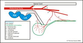
| + | [[Image:sumbsbladurtri.jpg|right|thumb|275px|<small><center>A schematic overview of the blood supply to the bladder and urethra</center></small>]] | ||
| + | ===Blood Supply=== | ||
| + | The bladder is supplied by cranial and a caudal vesical arteries. | ||
| + | |||
| + | =====Cranial Vesicular Artery===== | ||
| + | * Branch of the umbilical artery which branches directly off the internal iliac a. | ||
| + | * Often not present in the dog thanks to a great reduction of the umbilical artery after birth | ||
| + | * In cattle it is weak | ||
| + | * In pigs and horses it is relatively strong | ||
| + | |||
| + | =====Caudal Vesicular Artery===== | ||
| + | |||
| + | * Branch of the vaginal or prostatic artery | ||
| + | ** These in turn are branches of the internal pudendal artery which in turn is a branch of the internal iliac. | ||
| + | * Main supply to the bladder | ||
| + | |||
| + | =====Species Differences in the Origin of the Vaginal Artery===== | ||
| − | + | The description above relates to the bitch. However the vaginal artery branches directly off the internal iliac in some species. This is because the level of the division of the internal iliac a. into the internal pudendal a. and caudal gluteal a. varies. The division is more cranial in the dog and the horse so you have a short internal iliac and a long internal pudendal and the vaginal artery branches off the latter in these species. In the cow and pig however the reverse is true and division is far more caudal resulting in the vaginal artery actually being a branch of the internal iliac. | |
| − | + | ===Lymphatic Drainage=== | |
| − | + | *Iliosacral lymph nodes | |
| + | |||
| + | ==Alternative Anatomical Thinking== | ||
| + | |||
| + | Some people now think that rather than an internal sphincter which contracts to resist urine outflow there is an actual structure which is opened when its muscle bundles contract. This would mean in these species that urinary continence relies on the external sphincter. It is not certain as yet which structure is correct but this idea does concur with the recent demonstration that the proximal urethra plays a role in urine storage in the dog and goat. | ||
| + | |||
| + | {{Learning | ||
| + | |flashcards = [[Bladder Anatomy & Physiology Flashcards|Bladder Anatomy Flashcards]] | ||
| + | }} | ||
| − | + | {{OpenPages}} | |
| + | [[Category:Lower Urinary Tract - Anatomy & Physiology]][[Category:Bullet Points]] | ||
Revision as of 14:37, 5 July 2012
Introduction
- The bladder is where urine is stored before being expelled by the body through the micturition reflex.
- Without a bladder urinary continence would be impossible.
Anatomy
- It is a hollow, muscular organ
- It is divided for descriptive purposes into three parts
- Cranial Pole
- Intermediate body
- Caudal neck
- Its wall comprises a muscle layer covered in transitional epithelium.
- Its size and posistion are determined by how full it is.
- When empty the bladder wall is wrinkled and thicker
- It rests on the pubic bones
- It is entirely in the pelvis of the large species but partly enters the abdomen of the carnivores
- It is largely retroperitoneal in the larger species
- When full and distended the folds disappear and the wall appears thinner
- It then becomes intraperitoneal in the larger species
- When empty the bladder wall is wrinkled and thicker
- The trigone of the bladder gets its name as it looks like a triangle without a base.
- It is of clinical importance and is formed by the paired uteric folds
- These form by the orifice of the ureters
- It is visible even when the bladder is full
- The folds extend from the urethral opening to the neck of the bladder
- Where they merge to form the urethral crest
- It is believed to have increased sensitivty and is of differant embryological origin to the rest of tissue. More details can be found here
Muscles of the Bladder
The three muscular components of the bladder described below play a pivotal part in the micturition reflex.
Detrusor Muscle
This network of smooth muscle fibres lie in three sheets within the bladder wall and are supplied by both parasympathetic and sympathetic nerves. It is responsible for storage and expression of urine from the bladder.
Parasympathetic Supply - Detrusor Muscle
- S1-S3
- Synapse in pelvic plexus or bladder wall
- Pelvic nerves
- Innvervate the detrusor muscle
- Action - excitatory
- Function - empty bladder
Sympathetic Supply - Detrusor Muscle
- L1-L4
- Syanpse in caudal mesenteric ganglion - bladder wall
- Hypogastric nerve
- Receptor - beta 2
- Inhibitory action
- Allows bladder filling
Internal Urethral Sphincter
A thickening of the bladder musculature found at the neck of the bladder which is continuous with the detrusor and is therefore smooth muscle. However, unlike the detrusor its innervation is purely from sympathetic fibres.
Sympathetic Supply - Internal Urethral Sphincter
- L1-L4
- Synapse in caudal mesenteric ganglion
- Hypogastric nerve
- Receptor - alpha 1
- Excitatory action
- Function - retain urine through increased urethral tone
External Urethral Sphincter
This third component is more appropriately included in the anatomy section of the urethra.
The Ligaments of the Bladder
- Two lateral ligaments
- Insert in the dorsal abdominal wall
- Within them are the residual umbilical vessels which still have some patency and convey small amounts of blood to the cranial bladder
- They attach onto the bladder wall at the lateral vesical folds.
- Median ligament
- Connects the bladder to pelvic floor and linea alba
- Attaches into the bladder at the median vesical fold
- In the foetus this ligament supports the urachus.
Blood Supply, Innervation and Lymphatic Drainage
Blood Supply
The bladder is supplied by cranial and a caudal vesical arteries.
Cranial Vesicular Artery
- Branch of the umbilical artery which branches directly off the internal iliac a.
- Often not present in the dog thanks to a great reduction of the umbilical artery after birth
- In cattle it is weak
- In pigs and horses it is relatively strong
Caudal Vesicular Artery
- Branch of the vaginal or prostatic artery
- These in turn are branches of the internal pudendal artery which in turn is a branch of the internal iliac.
- Main supply to the bladder
Species Differences in the Origin of the Vaginal Artery
The description above relates to the bitch. However the vaginal artery branches directly off the internal iliac in some species. This is because the level of the division of the internal iliac a. into the internal pudendal a. and caudal gluteal a. varies. The division is more cranial in the dog and the horse so you have a short internal iliac and a long internal pudendal and the vaginal artery branches off the latter in these species. In the cow and pig however the reverse is true and division is far more caudal resulting in the vaginal artery actually being a branch of the internal iliac.
Lymphatic Drainage
- Iliosacral lymph nodes
Alternative Anatomical Thinking
Some people now think that rather than an internal sphincter which contracts to resist urine outflow there is an actual structure which is opened when its muscle bundles contract. This would mean in these species that urinary continence relies on the external sphincter. It is not certain as yet which structure is correct but this idea does concur with the recent demonstration that the proximal urethra plays a role in urine storage in the dog and goat.
| Urinary Bladder - Anatomy & Physiology Learning Resources | |
|---|---|
 Test your knowledge using flashcard type questions |
Bladder Anatomy Flashcards |
Error in widget FBRecommend: unable to write file /var/www/wikivet.net/extensions/Widgets/compiled_templates/wrt661eefaa0fa1c6_64273840 Error in widget google+: unable to write file /var/www/wikivet.net/extensions/Widgets/compiled_templates/wrt661eefaa15b297_83721353 Error in widget TwitterTweet: unable to write file /var/www/wikivet.net/extensions/Widgets/compiled_templates/wrt661eefaa1b4318_72590522
|
| WikiVet® Introduction - Help WikiVet - Report a Problem |


