File:Chicken Egg schematic.jpg
Jump to navigation
Jump to search
No higher resolution available.
Chicken_Egg_schematic.jpg (283 × 351 pixels, file size: 51 KB, MIME type: image/jpeg)
Summary
| Description |
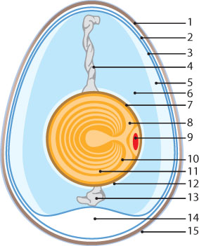
1. Eggshell 2. Outer membrane 3. Inner membrane 4. Chalaza 5. Exterior albumen (outer thin albumen) 6. Middle albumen (inner thick albumen) 7. Vitelline membrane 8. Nucleus 9. Germinal disk (blastoderm) 10. Yellow yolk 11. White yolk 12. Internal albumen 13. Chalaza 14. Air cell 15. Cuticle |
|---|---|
| Date |
2008 |
| Source |
RVC |
| Author |
unknown |
| Permission (Reusing this file) |
See below |
Licensing
| This file is licensed under the Creative Commons Attribution Non-Commercial & No Derivative Works License |
File history
Click on a date/time to view the file as it appeared at that time.
| Date/Time | Thumbnail | Dimensions | User | Comment | |
|---|---|---|---|---|---|
| current | 07:32, 31 July 2008 | | 283 × 351 (51 KB) | Lwilkie (talk | contribs) | 1. Eggshell 2. Outer membrane 3. Inner membrane 4. Chalaza 5. Exterior albumen (outer thin albumen) 6. Middle albumen (inner thick albumen) 7. Vitelline membrane 8. Nucleus 9. Germinal disk (blastoderm) 10. Yellow yolk 11. White yolk 12. Inter |
You cannot overwrite this file.
File usage
The following file is a duplicate of this file (more details):
- File:Ei1.jpg from Wikimedia Commons
The following 2 pages use this file: