Difference between revisions of "Adenoma"
| Line 1: | Line 1: | ||
| + | {{OpenPagesTop}} | ||
==Introduction== | ==Introduction== | ||
[[Image:dogpap1.gif|right|thumb|100px|<small><center>Oral Papilloma Neoplasia in Dog (Courtesy of Alun Williams (RVC))</center></small>]] | [[Image:dogpap1.gif|right|thumb|100px|<small><center>Oral Papilloma Neoplasia in Dog (Courtesy of Alun Williams (RVC))</center></small>]] | ||
| Line 85: | Line 86: | ||
{{review}} | {{review}} | ||
| + | |||
| + | {{OpenPages}} | ||
[[Category:Expert Review]] | [[Category:Expert Review]] | ||
[[Category:Oropharynx - Pathology]] | [[Category:Oropharynx - Pathology]] | ||
Revision as of 15:38, 5 July 2012
Introduction
An adenoma is a benign epithelial tumour arising in the epithelium of the mucosa (stomach and intestines), glands (endocrine and exocrine) and ducts.
Adenomas observed in veterinary species include:
Perianal Adenoma
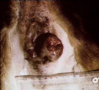
These tumours, also called hepatoid gland tumours, arise from the solid, modified sebaceous circumanal glands. They are the third most common tumour in intact male dogs, and arise more frequently in older dogs.
The tumour is under hormonal control. Hepatoid glands are also found at the tail head, prepuce and other skin sites, and tumours can also arise from there.
Clinical features
Adenomas occur alone or in number, as round, well-differentiated, freely-movable masses. Tumours can become ulcerated and secondarily infected. There can be signs of perianal pain and tenesmus.
Diagnosis
Cytology of the mass will reveal large hepatoid cells with a round, central nuclei, multiple nucleoli, and an abundant cytoplasm. There may be concurrent inflammation or haemorrhage. Cytology cannot distinguish adenomas from adenocarcinomas, and further investigations should be carried out if malignancy is suspected.
Treatment
Castration is the treatment of choice and 95% of tumours will regress. Administration of oestrogens or anti-androgens can also be considered, but side-effects of those hormones should not be forgotten. Surgical removal of the tumour may be necessary if it is large, or in females.
Sweat Gland Adenoma
This is a tumour of the apocrine sweat gland and is rare in dogs and cats. It can be difficult to differentiate from an adenocarcinoma, and immunohistochemistry has been used for this purpose.
Adenomas rarely ulcerate, are associated with little local inflammation and have a cystic feel on palpation.
They occur most commonly in older dogs and cats, and are usually restricted to the head.
Wide surgical excision usually carries a good prognosis.
Ceruminous Gland Adenoma
This occurs with some frequency in dogs and cats, and is thought to be linked to the presence of long-standing otitis externa, leading to increased glandular dysplasia.
These tumours usually occur in older animals, and conservative local resection is usually sufficient to manage them.
Sebaceous Gland Adenoma
These are common in older dogs and cats and are usually distinctly wart-like or cauliflower-like in appearance.
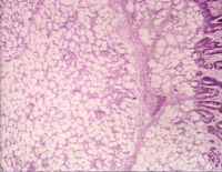
Histopathology shows large mature sebaceous lobules with increased numbers of basaloid epithelial cells and a low mitotic activity.
The prognosis is good with surgical resection.
Salivary Gland Adenoma
This tumour is rare in animals, and the malignant adenocarcinoma is much more common.
Mammary Gland Adenoma
This is a benign tumour which is quite common in cats and dogs.
Find out more information on mammary tumours.
Intestinal Adenoma
Intestinal adenomas are found in both the small and large intestines. Intestinal adenomas usually grow into the lumen and can be called adenomatous polyps.
Depending on the type of the insertion base, the adenoma may be pedunculated with a long stalk, or sessile with a broad base. This influences the method of resection and the rate of recurrence, as pedunculated tumours are much more easily removed.
Hepatic Adenoma
It is seen mostly in sheep and cattle and usually presents as a single, pale, soft, often large nodule, which is well demarcated from adjacent tissue, often with a noticeable capsule. The tissue has a normal hepatocytic appearance. No portal tracts can be seen within the mass and a capsule surrounds the growth.
Cholangiocellular Adenoma
Also called biliary adenoma, it is very rare but has been reported in dogs and cats. It shows an expansive growth and consists of slightly dilated, occasionally cystic structures, lined with cuboidal or flattened, well differentiated biliary epithelium.
Pancreatic Adenoma
Image of multifocal pancreatic adenoma in a dog from Cornell Veterinary Medicine Adenoma of the exocrine (zymogen) cells of the pancreas is known in several species and is recognised by its ductal or acinar pattern of cells, with an expanding growth pattern and complete encapsulation. Cystic spaces may be created by the tumour cells, which may also project in a papillary pattern into the lumen of the cysts.
Hyperplastic nodules may be present in the pancreas of older animals. They are usually less well encapsulated than adenomas, but may be difficult to distinguish with certainty. They are usually multiple.
| Adenoma Learning Resources | |
|---|---|
 Test your knowledge using flashcard type questions |
Cytology Q&A 07 |
References
Withrow, S. (2001) Small animal clinical oncology Elsevier Health Sciences
Morrison, W. (2002) Cancer in dogs and cats: medical and surgical management Teton NewMedia
Carlyle Jones, T. (1997) Veterinary pathology Wiley-Blackwell
Cheville, N. (1999) Introduction to veterinary pathology Wiley-Blackwell
| This article has been peer reviewed but is awaiting expert review. If you would like to help with this, please see more information about expert reviewing. |
Error in widget FBRecommend: unable to write file /var/www/wikivet.net/extensions/Widgets/compiled_templates/wrt672419a759cf79_85263253 Error in widget google+: unable to write file /var/www/wikivet.net/extensions/Widgets/compiled_templates/wrt672419a763b235_65768661 Error in widget TwitterTweet: unable to write file /var/www/wikivet.net/extensions/Widgets/compiled_templates/wrt672419a767e7c4_90306435
|
| WikiVet® Introduction - Help WikiVet - Report a Problem |


