Difference between revisions of "Exploratory Laparotomy - Donkey"
| Line 29: | Line 29: | ||
* paramedian approach for cryptorchidism | * paramedian approach for cryptorchidism | ||
* ventral abdominal approach for colic and caesarean operations | * ventral abdominal approach for colic and caesarean operations | ||
| + | |||
| + | <big> | ||
[[Pyometra - Donkey|'''Pyometra''']] | [[Pyometra - Donkey|'''Pyometra''']] | ||
| + | [[Ovariectomy - Donkey|'''Ovariectomy''']] | ||
| − | |||
| − | |||
| − | |||
The indications for, and treatment of, bladder stones and cryptorchidism are similar to those in the horse. | The indications for, and treatment of, bladder stones and cryptorchidism are similar to those in the horse. | ||
Revision as of 00:03, 20 February 2010
| This article has been peer reviewed but is awaiting expert review. If you would like to help with this, please see more information about expert reviewing. |
Indications for Exploratory Laparotomy
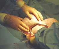
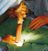
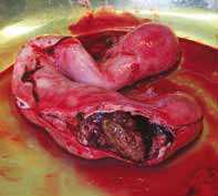
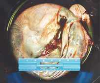
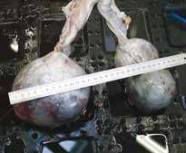
Exploratory laparotomy may be required in the donkey, as in the horse, for emergency or elective purposes, such as surgical colic, caesareans, and uro-genital surgery.
As discussed, colic in the donkey, especially impaction colic, may present with more subtle clinical signs than in a horse. However, acute signs of pain with nasogastric reflux and distended viscera are occasionally seen, and may indicate prompt surgical intervention. It is important to make an assessment of triglyceride levels and pancreatic enzymes as speedily as possible, as concurrent hyperlipaemia and/or pancreatitis will reduce the prognosis even if recognised and treated. Pancreatitis can present with signs of peracute pain in the anterior abdomen, which may be exacerbated by external ballotment behind the ventral sternum. In addition, peritoneal tap samples may show high levels of neutrophils, amylase and lipase.
Examination
The examination of the donkey with suspected surgical colic may be hampered due to the small size of the patient. A rectal examination is nearly always possible even if sedation is required. If this is truly impossible, ultrasound examination of the abdomen externally should be performed to assess viscera size, position and motility.
A peritoneal tap provides useful information; however most UK donkeys carry considerable fat deposits above the linea alba. If ultrasound is available, the depth of the peritoneal fat may be measured and an appropriate needle or (more safely) a teat cannula used.
In aged donkeys, a good assessment of dental disorders and possible chronic hoof disease should be made before surgery is contemplated. Our records show that internal neoplasia is also a significant complicating factor in geriatric donkeys. This should be taken into account and discussed with the donkey owner, especially if there has been a history of progressive weight loss, or unexplained changes in haematological/biochemical parameters. Despite all the warnings, donkeys have been successfully operated on for correctable surgical lesions, for example, small intestine strangulation and large colon displacements.
Urogenital surgery
Surgical correction of urogenital problems has been used at The Donkey Sanctuary for the following conditions: caesarean section, ovariohysterectomy for pyometra, ovary removal, bladder stone removal, and cryptorchidism. The approach required will depend on the surgery to be performed.
We have used
- flank laparatomy for unilateral ovarian removal
- paramedian approach for cryptorchidism
- ventral abdominal approach for colic and caesarean operations
The indications for, and treatment of, bladder stones and cryptorchidism are similar to those in the horse.
Caesarean section
The indications are as for the horse (e.g. foetal oversize, dystocia) and, in most cases, a ventral midline approach is used as in the horse. Many donkeys are poorly supervised around foaling and may be presented for dystocia in a state of endotoxic shock. In a paper by Bourassi and Kay (2006), the results of standing left flank approach to caesarean section under sedation and local anaesthesia is presented. Although the sample size was small, the authors comment that the technique may provide a safe, low cost and viable alternative to conventional mid-line laparotomy. In addition, the same authors suggest that donkeys carrying hinny foals may be more susceptible to dystocia due to poorer regulation of foetal growth.
References
- Thiemann, A. (2008) Surgery In Svendsen, E.D., Duncan, J. and Hadrill, D. (2008) The Professional Handbook of the Donkey, 4th edition, Whittet Books, Chapter 16
- Thiemann, A.K., Makhambeni, M.M.S. (2006). ‘Five cases of ovariohysterectomy in the donkey’. 'Proceedings of the 9th congress of the W.E.V.A. pp 487-489.
- Abd El-Karim, R.(1995). ‘Two cases of rectal prolapse in the donkey’.Equine Veterinary Education 7 (1). pp 12-14.
- Bell, N.J., Thomas, S. (2001). ‘Use of sterile maggots to treat panniculitis in an aged donkey’. Veterinary Record 149. pp 768-770.
- Bouayad, H., Rifai, S., Kay, R.S., Knottenbelt, D.C., and Smith, M. (2006). ‘Bifid tongue and Mandibular cleft in three mule foals’. Proceedings of the 9th Congress of the W.E.V.A. pp 334-346.
- Bourassi, M., Kay, G. (2006). ‘Dystocia in donkeys carrying mule foals in Morocco: an evaluation of 32 cases’. Proceedings of the 9th Congress of the W.E.V.A.
- Farmand, M., Stohler, T. (1990). ‘The median cleft of the lower lip and mandible and its surgical correction in a donkey’. Equine Veterinary Journal 22 (4). pp 298-301.
- Kay, G. (2005). ‘Sialolithiasis in donkeys’. In: Veterinary care of donkeys. Matthews, N.S., and Taylor, T.S. (eds). International Veterinary Information Service. www.pubmedcentral.nih.gov/articlerenderfcgi?articl=14449901
- Misk, N.A., Nigam, M.V. (1984). ‘Sialolith in a Donkey’. Equine Practice 6(4). pp 49-50.
- Thiemann, A.K. (2003). ‘Treatment of a deep injection abscess using sterile maggots in a donkey’. World Wide Wounds website. November 2003 (www.worldwidewounds.com)
|
|