Meninges - Anatomy & Physiology
Jump to navigation
Jump to search
| This article is still under construction. |

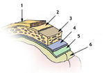
Diagram of the meninges. Sourced from WikiMedia Commons, attributed to http://training.seer.cancer.gov/module_anatomy/unit5_3_nerve_org1_cns.html
- The CNS is surrounded by several layers of tissue.
- Bone layer
- E.g. the skull and the vertebrae.
- Dura-arachnoid layer.
- Composed of the dura mater and arachnoid layer in close opposition.
- Adhered to the skull in the calvaria.
- Pia mater.
- Bone layer
- The epidural space lies between the bone layer (skull or vertebrae) and the dura-arachnoid layer.
- Local anaesthetic is injected here in epidural anaesthesia.
- E.g. when calving cows.
- Local anaesthetic is injected here in epidural anaesthesia.
- The sub-arachnoid space lies between the arachnoid layer and the pia mater.
- CSF filled.
- This is where myelographic contrast medium is injected.