Central Nervous System - Histology
Introduction
The Central Nervous System (CNS) is composed of the brain and the spinal cord and it is from the CNS that the Peripheral Nervous System (PNS) is derived. This page is specifically focussed on the histologic appearence of both the brain and spinal cord and therefore the anatomy of both structures will not be provided in depth. Instead, links to the relevant anatomy and physiology pages for each structure will be given below.
Anatomy & Physiology Links
Please use the links below to familiarise yourself with the anatomy and physiology of the different aspects of the CNS as an aid to utilising and understanding the CNS histology images below;
CNS Development
Forebrain
Midbrain
Hindbrain
Cranial Nerves
Spinal Cord
Histology of the Spinal Cord
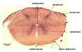
The spinal cord is composed to two discrete parts; the white matter which is the outer part of the cord and the grey matter which is the inner portions of the cord. The white matter is given this name due to its appearance in unfixed histological specimens in which the white nature of the tissue is caused by the myelination of ascending and descending nerve fibres. The grey matter is also named after its unfixed histological appearance and contains the cell bodies of neurons as well as nerve fibres.
Within the spinal cord the grey matter forms an H-shape where the ventral horns of the H are broader than the dorsal horns. The grey matter shape has also been likened to that of a butterfly. The grey matter also has a histologically visible central canal running through it. The ventral horns of the grey matter contain the cell bodies of motor neurones whilst the dorsal horns contain sensory neurons where the cell bodies are found in the dorsal root ganglia. Please see sensory pathways for further information on the composition of nerve fibres within the spinal cord. The relative size of the grey matter is dependant on the number of motor cells related to controlling limbs and therefore the size varies along the length of the spine. Around the areas of the fore and hindlimbs the grey matter is considerably larger.

The above image shows a complete cross-sectional histology of a spinal cord.


Rabbit Spinal Cord

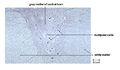
Spinal cord TS section
The main cell type here is neuroglial cells.
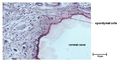
Spinal cord TS section
This section shows well the large multipolar cells within the ventral horn.
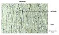
Spinal cord TS section
Histology of the Brain


Cerebrum section
Cerebellum section
Spinal Cord - © John Bredl
Spinal Cord 2 - © John Bredl
Spinal Cord 3 - © John Bredl
Cerebrum - © John Bredl
Cerebellum - © John Bredl


| This article is still under construction. |