Difference between revisions of "Female Reproduction - Donkey"
| Line 7: | Line 7: | ||
'''[[Oestrus Cycle - Donkey|Oestrous cycle]] | '''[[Oestrus Cycle - Donkey|Oestrous cycle]] | ||
| + | |||
| + | '''[[Pregnancy - Donkey|Pregnancy]] | ||
</big> | </big> | ||
| − | |||
| − | |||
| − | |||
| − | |||
| − | |||
| − | |||
| − | |||
| − | |||
| − | |||
| − | |||
| − | |||
| − | |||
| − | |||
| − | |||
| − | |||
| − | |||
| − | |||
| − | |||
| − | |||
| − | |||
| − | |||
| − | |||
| − | |||
| − | |||
| − | |||
| − | |||
| − | |||
==Parturition and ''post-partum''== | ==Parturition and ''post-partum''== | ||
[[Image:Late term jenny donkey.jpg|right|thumb|300px|<small><center>Late term pregnant female showing mammary development and ventral oedema. Although this development takes place primarily in the last 48 hours of pregnancy, it may be present in some females up to two weeks before parturition. (Image courtesy of [http://drupal.thedonkeysanctuary.org.uk The Donkey Sanctuary])</center></small>]] | [[Image:Late term jenny donkey.jpg|right|thumb|300px|<small><center>Late term pregnant female showing mammary development and ventral oedema. Although this development takes place primarily in the last 48 hours of pregnancy, it may be present in some females up to two weeks before parturition. (Image courtesy of [http://drupal.thedonkeysanctuary.org.uk The Donkey Sanctuary])</center></small>]] | ||
Revision as of 22:00, 22 February 2010
| This article has been peer reviewed but is awaiting expert review. If you would like to help with this, please see more information about expert reviewing. |
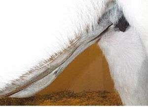
Female reproductive anatomy peculiarities
Parturition and post-partum

The length of pregnancy is 365 to 376 days but extreme variations ranging from 340 to 395 days have been reported (Kohli et al, 1957; Tibary and Bakkoury, 1994; Macusoe et al, 2004). Male foetuses are carried on average one day longer.
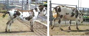
Prediction of foaling is based on udder development and waxing, which seems to reliably start 24 to 48 hours before foaling. The calcium concentration in mammary secretions increases steadily in the last week of pregnancy and surpasses 500 ppm about 24 hours before foaling.
Parturition presents in the classical three stages:


- Stage 1: (preparation for labour) Characterized by increased restlessness, isolation from herd mates and frequent alternation between standing and lying down.
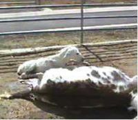
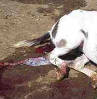
- Stage 2: (expulsion of the foetus) starts with the rupture of the allantochorion and appearance of the amniotic sac at the vulva. This is usually completed in 10 to 30 minutes with the female in lateral recumbency.
- Stage 3: (expulsion of placenta) usually completed in one hour but may take up to six hours. Possible retention of the placenta should be closely monitored and treated if the placenta is not passed by six hours. Treatment with oxytocin intravenously (in lactate ringer) or intramuscularly is effective. However, jennies with retained placenta should also be monitored for hypocalcaemia.
Parturition can be induced by the administration of small doses of oxytocin. Criteria for induction of parturition are similar to those described for the mare, that is, pregnancy length >340 days, evidence of mammary development and waxing, change in pre-colostrum electrolyte and no abnormalities (Pashen, 1980).
Donkey dystocia cases have been reported, but their true incidence and nature is not known. Malformations such as schistosomus reflexus and foetal ankylosis have been reported as causes of dystocia (Dubbin et al, 1990). In miniature donkeys, dystocia may result following abortion due to foetal malformation. In equines, the uterus should be able to regulate foetal growth and reduce the rate of dystocia due to foetal-maternal disproportion. However, an increased rate of dystocia is commonly feared when jennies are bred to stallions.
Dystocia risks are increased in miniature donkeys because of the domed large forehead of some foals. Relief of dystocia is best accomplished under anaesthesia, with the jenny’s hindquarters suspended. Caesarean section is preferable to foetotomy. Long obstetrical manipulations have been associated with vaginal adhesions.
Uterine involution is still poorly studied in asinine species. One study on Catalonian jennies reported that gross uterine involution is completed by 22.5 ± 1.7 days post-partum (range of 18 to 27 days) (Dadarwal et al, 2004). Vaginal discharge (clear odourless lochia) was present for up to seven days and intrauterine fluid was detected by ultrasound for up to 18 days in some females.
Foal heat occurs 8.6 days after foaling, but periods of 2 to 69 days have been reported. Dardawal et al (2004) found that, on the day of parturition, several 10 to 15 mm follicles are present and by 5 to 12 days post-partum at least one follicle has reached 25 mm in diameter. Behavioural oestrus seems to be less pronounced with the first post-partum ovulation.
Most jennies will ovulate between 13 to 17 days post-partum. Extended periods of follicular activity without ovulation may be observed in some females. The interaction between post-partum cyclicity, body condition and lactation has not been investigated. In a study from 1957 in India, the mean interval from parturition to fertilization was found to be 162.5 ± 6.7 days (Kohli et al, 1957).
There are very few studies on nutritional requirements for optimal fertility, pregnancy needs and lactation. In one study, jennies lost abut 1% of their body weight at foaling and a further 0.5 to 1% during lactation (Eley and French, 1994).
Donkey neonate survival is often described as a major problem in developing countries. This high mortality may be due to poor rearing conditions (post-partum care) as well as poor colostral protection. The effect of nutrition and management on foal growth and survival has not been fully studied.
In a free-ranging population, jennies may continue to nurse their foals until ten months of age (French, 1998). Feed supplementation along with anthelmintic treatment during late pregnancy and lactation has been associated with better survival of foals in Ethiopia (Mengistu et al, 2005). This may be due to increased mammary function and prevention of failure of passive transfer.
References
- Tibary, A., Sghiri, A. & Bakkoury, M. (2008) Reproduction In Svendsen, E.D., Duncan, J. and Hadrill, D. (2008) The Professional Handbook of the Donkey, 4th edition, Whittet Books, Chapter 17
- Blanchard, T.L., Taylor, T.S. (2005). ‘Oestrous Cycle Characteristics of Donkeys with Emphasis on Standard and Mammoth Donkeys’. in: Veterinary Care of Donkeys. N.S. Matthews and T.S. Taylor (eds).
- Blanchard, T.L., Taylor, T.S., and Love, C.L. (1999). ‘Estrous cycle characteristics and response to estrus synchronization in mammoth asses (Equus asinus americanus)’. Theriogenology 52. pp 827-834.
- Carluccio, A., Villani, M., Contri, A., Tosi, U., and Battocchio, M. (2004). ‘Preliminary study on some seminal and testicular morphometric characteristics in Martina Franca jackass’. Ippologia 15. pp 23-26.
- Carluccio, A., Villani, M., Contri, A., Tosi, U., and Veronesi, M.C. (2004).‘Ultrasonographic evaluation of the early pregnancy in Martina Franca jennies’. Ippologia 16. pp 31-35.
- Dadarwal, D., Tandon, S.N., Purohit, G.N., and Pareek, P.K. (2004). ‘Ultrasonographic evaluation of uterine involution and post-partum follicular dynamics of French Jennies (Equus asinus)’. Theriogenology 62. pp 257-264.
- Dubbin, E.S., Welker, F., Veit, H., Modransky, P.D., Talley, M.R., Vandeplassche, M., and Salah-Osman-Idris, M.B. (1990). ‘Dystocia attributable to a fetal monster resembling schistosomus reflexus in a donkey’. Journal of the American Veterinary Association 197. pp 605-607.
- Eley, J.L., French, J.M. (1994). ‘Bodyweight changes in pregnant and lactating donkey mares and their foals’. Veterinary Record 134. p 627.
- Fielding, D. (1988). ‘Reproductive characteristics of the jenny donkey - Equus asinus: a review’. Tropical Animal Health and Production 20. pp 161-166.
- French, J.M.(1998). ‘Mother-offspring relationships in donkeys’. Applied Animal Behaviour Science 60. pp 253-258.
- Ginther, O.J., Scrabam, S.T., and Bergfelt, D.R. (1987). ‘Reproductive seasonality of the jenny’. Theriogenology 27. pp 587-592.
- Henry, M., Figueiredo, A.E.F., Palhares, M.S., and Coryn, M. (1987). ‘Clinical and endocrine aspects of the estrous cycle in donkeys (Equus asinus)’. Journal of Reproduction and Fertility Suppl.35. pp 297-303.
- Kohli, M.L., Suri, K.R. (1957). ‘Studies on reproductive efficiency in donkey mares’. Indian J Vet. Sci. 27. pp 133-138.
- Kohli, M.L., Suri, K.R., and Chatter, J.I.A. (1957). ‘Studies on the gestation length and breeding age in donkey mares’. Indian Vet J. 34, pp 344-348.
- Lemma, A., Bekana, M., Schwartz, H.J., and Hildebrant, T. (2006). ‘Ultrasonographic study of ovarian activities in the tropical jenny during the seasons of high and low sexual activity’. Journal of Equine Veterinary Science 26. pp 18-22.
- Lemma, A., Bekana, M. , Schwartz, H.J., and Hildebrant, T. (2006) ‘The effect of body condition on ovarian activity of free ranging tropical jennies (Equus asinus)’. Journal of Veterinary Medicine Series A- Physiology and Pathology Clinical Medicine 53. pp 1-4.
- Lun, S., He, C., Zhang, Y., Chen, Z., Hu, W., Lu, S., Jia, W., and Zhang, Q. (1998). ‘Preliminary study on development of follicles and formation of corpus luteum of the jenny and mare by ultrasonography’. Acta Veterinaria et Zootechnica Sinica 29. pp 419-425.
- Mancuso, R., Torrisi, C., and Catone, G. (2004). ‘Experiences in the management of the reproductive activity of dairy jennie-ass’. Ippologia 15. pp 5-9.
- Meira, C.,Ferreira, J.C.P., Papa, F.O., and Henry, M. (1995). ‘Study of the estrous cycle in donkeys (Equus asinus) using ultrasonography and plasma progesterone concentrations’. Biol Reprod Monograph 1. pp 403-410.
- Meira, C., Ferreira, J.C.P. , Papa, F.O., and Henry, M. (1998). ‘Ovarian activity and plasma concentrations of progesterone and estradiol during pregnancy in jennies’. Theriogenology 49. pp 1465-1473.
- Meira, C., Ferreira, J.C.P., Papa, F.O., and Henry, M. (1998). ‘Ultrasonographic evaluation of the conceptus from 10 days to 60 of pregnancy in jennies’. Theriogenology 49. pp 1475-1482.
- Mengistu, A., Smith, D.G., Yoseph, S., Nega,T., Zewdie, W. , Kassahun, W.G., Taye, B., and Firew, T. (2005). ‘The effect of providing feed supplementation and anthelmintic to donkeys during late pregnancy and lactation on live weight and survival of dams and their foals in central Ethiopia’. Tropical Animal Health and Production 37 (supplement 1). pp 21-33.
- Orozco Hernandez, J.L., Escobar Medina, F.J., and De La Colina Flores, F. (1992). ‘Ovarian activity in the mare and the female donkey during days with less light’. Veterinaria Mexico 23. pp 47-50.
- Pashen, R.L. (1980). ‘Low doses of oxytocin can induce foaling at term’. Eq. Vet. J. 12. pp 85-87.
- Purdy, S.R. (2005a). ‘Artificial Insemination for Miniature Donkeys’. in: Veterinary Care of Donkeys. N.S. Matthews and T.S. Taylor (eds). International Veterinary Information Service, Ithaca NY (www.ivis.org), 20 Apr, 2005; A2924.0405.
- Purdy, S.R. (2005b). ‘Ultrasound examination of the female miniature donkey reproductive tract’. in: Veterinary Care of Donkeys. N.S. Matthews and T.S. Taylor (eds). International Veterinary information Service, Ithaca NY (www.ivis.org), last updated: 11 May 2005; A2925.0505.
- Tibary, A. (2004). ‘Reproductive patterns in donkeys and minature horses’. Proceedings of the North American Veterinary Conference, Orlando, Florida, 17-21 January. pp 231-233, 2004.
- Tibary, A., Bakkoury, M.(eds). (1994). ‘Particularités de la reproduction chez les autres espèces équines’. Reproduction équine, Tome I: La jument. Actes Editions 1994. pp 385-400.
- Vandeplassche, G.M., Wesson, J.A., and Ginther, O.J. (1981). ‘Behavioral, follicular and gonadotropin changes during estrous cycle in donkeys’. Theriogenology 16. pp 239-249.
- Vendramini, O.K., Guintard, C., Moreau, J., and Tainturier, D. (1998). ‘Cervix conformation: a first anatomical approach in Baudet du Poitou jenny asses’. Animal Science 66. pp 741-744.
|
|
|
|