Difference between revisions of "Lungs - Anatomy & Physiology"
Jump to navigation
Jump to search
| Line 56: | Line 56: | ||
Image:Bronchuslowpower2.jpg|<p>'''Bronchus'''</P><sup>©RVC 2008</sup> | Image:Bronchuslowpower2.jpg|<p>'''Bronchus'''</P><sup>©RVC 2008</sup> | ||
Image:Alveoli.jpg|<p>'''Alveoli'''</P><sup>©RVC 2008</sup> | Image:Alveoli.jpg|<p>'''Alveoli'''</P><sup>©RVC 2008</sup> | ||
| + | Image:Alveoli2.jpg|<p>'''Alveoli'''</P><sup>©RVC 2008</sup> | ||
</gallery></center> | </gallery></center> | ||
Revision as of 14:46, 12 August 2008
|
|

Introduction

The lungs are the site for gaseous exchange, and are situated within the thoracic cavity. They occupy approximatley 5% of the body volume in mammals when relaxed, but generally have no fixed size or shape since their volume is constantly changing with the processes of inspiration and expiration.
The lungs, along with the larynx and trachea, develop from a ventral respiratory tract. After separation from the developing oesophagus, two lung buds develop, which undergo divisions as they grow, forming the beginnings of the bronchial tree. This process is not completed by birth.
Structure
- The left and right lungs lie within their pleural sac and are only attached by their roots, to the mediastinum, so they are
- The right lung is always larger than the left, due to the positioning of the heart. The apex of the lungs is the cranial point.
- In most species the lungs are divided into lobes by the bronchial tree:
- Left Lung = Cranial and Caudal lobes.
- Right Lung = Cranial, Caudal, Middle and Accessory lobes. The cranial lobe is further divided by an external fissure.
- The bulk of the lung consists of bronchi, blood vessels and connective tissue. The terminal bronchioles have alveoli scattered along their length, and are continued by alveolar ducts, alveolar sacs and finally alveoli.
File:Routeofairthroughrespiratorysystem.jpg
Schematic Diagram showing the route air takes through the respiratory system
Function
- Gaseous Exchange
Vasculature
- The 'Pulmonary Arteries follow the bronchi while the pulmonary veins sometimes run separately.
- Bronchial arteries from the Aorta supply the bronchi, and Bronchial veins may drain this blood to the right atrium via the Azygous Vein.
Innervation
- Nervous supply to the lung is via the Pulmonary Plexus within the mediastinum.
Lymphatics
- Lymph drains to the Tracheobronchial and Mediastinal lymph nodes.
Histology
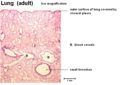
Lung (adult)
©RVC 2008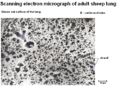
SEM of Adult Sheep Lung
©RVC 2008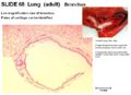
Bronchus
©RVC 2008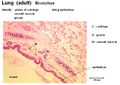
Bronchus
©RVC 2008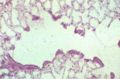
Alveoli
©RVC 2008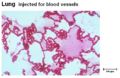
Alveoli
©RVC 2008


Species Differences
- Externally the lungs of the Horse show almost no lobation. Internally the right lung of the horse lacks a Middle Lobe.
- The lungs of Ruminants and Pigs are obviously lobed.
- The fissures between lobes are deeper in the dog and cat lung compared to other species.
Links
References
- Dyce, K.M., Sack, W.O. and Wensing, C.J.G. (2002) Textbook of Veterinary Anatomy. 3rd ed. Philadelphia: Saunders.