Difference between revisions of "Lymph Nodes - Anatomy & Physiology"
Fiorecastro (talk | contribs) |
|||
| (46 intermediate revisions by 6 users not shown) | |||
| Line 1: | Line 1: | ||
| − | |||
| − | |||
| − | |||
| − | |||
| − | |||
| − | |||
| − | |||
| − | |||
| − | |||
| − | |||
| − | |||
| − | |||
| − | |||
| − | |||
| − | |||
| + | [[Image:Lymph Node positions.jpg|thumb|right|250px|Positions of the lymph nodes (dog) - Copyright B.Catchpole RVC]] | ||
| + | [[Image:Aspinall Slide11.JPG|thumb|right|250px|<small>Image from [http://www.elsevierhealth.co.uk/veterinary-nursing/spe-60136/ Aspinall, The Complete Textbook of Veterinary Nursing], Elsevier Health Sciences, ''All rights reserved''</small>]] | ||
==Introduction== | ==Introduction== | ||
''For haemolymph nodes click [[Haemolymph Nodes - Anatomy & Physiology|here]]'' | ''For haemolymph nodes click [[Haemolymph Nodes - Anatomy & Physiology|here]]'' | ||
| − | [[Image:LH Lymph Node Diagram.jpg|thumb| | + | [[Image:LH Lymph Node Diagram.jpg|thumb|250px|right|<p>'''Diagram'''</p>]] |
| − | <p>Part of the [[Lymphatic System - Anatomy & Physiology|lymphatic system]], the body contains hundreds of lymph nodes of varying size (1-20mm) and these are located along the routes of [[Lymphatic Vessels - Anatomy & Physiology|lymphatic vessels]]. They are found throughout the body but are more concentrated in the axilla, groin and mesenteries. Lymph nodes act as a filter for the lymph removing antigens and releasing immune-competent cells and immunoglobulins.</p> | + | <p>Part of the [[Lymphatic System Overview - Anatomy & Physiology|lymphatic system]], the body contains hundreds of lymph nodes of varying size (1-20mm) and these are located along the routes of [[Lymphatic Vessels - Anatomy & Physiology|lymphatic vessels]]. They are found throughout the body but are more concentrated in the axilla, groin and mesenteries. Lymph nodes act as a filter for the lymph removing antigens and releasing immune-competent cells and immunoglobulins.</p> |
==Development== | ==Development== | ||
| − | <p>Lymph nodes develop from lateral plate mesoderm in paired sacs from [[Lymphatic Vessels - Anatomy & Physiology|lymphatic vessels]]. These sacs undergo remodelling and endothelial and mesenchymal outgrowths form the meshwork of channels and spaces that produces the cortex-medulla structure. Lymphocytes then populate the cortex and medulla. The | + | <p>Lymph nodes develop from lateral plate mesoderm in paired sacs from [[Lymphatic Vessels - Anatomy & Physiology|lymphatic vessels]]. These sacs undergo remodelling, and endothelial and mesenchymal outgrowths form the meshwork of channels and spaces that produces the cortex-medulla structure. Lymphocytes then populate the cortex and medulla. The subcapular sinus is a remainder of the [[Lymphatic Vessels - Anatomy & Physiology|lymphatic vessel]].</p> |
| + | |||
==Structure== | ==Structure== | ||
| − | [[Image:LH_Lymph_Node_Follicles_Histology.jpg|thumb| | + | [[Image:Aspinall Slide10.JPG|thumb|right|250px|<small>Image from [http://www.elsevierhealth.co.uk/veterinary-nursing/spe-60136/ Aspinall, The Complete Textbook of Veterinary Nursing], Elsevier Health Sciences, ''All rights reserved''</small>]] |
| − | [[Image:LH_Lymph_Node_Follicle_Histology.jpg|thumb| | + | [[Image:LH_Lymph_Node_Follicles_Histology.jpg|thumb|250px|right|<p>'''Primary & secondary follicles'''</p><sup>©Nottingham Uni 2008</sup>]] |
| + | [[Image:LH_Lymph_Node_Follicle_Histology.jpg|thumb|250px|right|<p>'''Secondary Follicle'''</p><sup>©RVC 2008</sup>]] | ||
===General=== | ===General=== | ||
| − | <p>Grossly the lymph nodes are round or bean shaped and have an outer cortex and an inner medulla. Microscopically the nodes have | + | <p>Grossly the lymph nodes are round or bean shaped and have an outer cortex and an inner medulla. Microscopically the nodes have follicles, paracortical zones and medullary cords and sinuses. At the hilum the medulla is present on the outer part of the node. Lymph nodes are located in series with [[Lymphatic Vessels - Anatomy & Physiology|lymphatic vessels]]. Afferent vessels enter the node on its convex side and efferent vessels exit on its concave side.</p> |
===Capsule and reticular framework=== | ===Capsule and reticular framework=== | ||
| − | <p> The nodes are surrounded | + | <p> The nodes are surrounded by a fibrous capsule that extends into the node as trabeculae, which provide an overall framework. Below the capsule is the subcapsular sinus. The nodes' parenchyma contain a fine network of reticular fibres and reticular cells. Reticular cells provide "scaffolding" for other cells as well as expressing surface complexes and substance to attract [[Lymphocytes#T cells|T cells]], [[Lymphocytes#B cells|B cells]] and [[T cell differentiation#Dendritic Cells|dendritic cells]]. |
| − | The cortex has aggregations of [[Lymphocytes | + | The cortex has aggregations of [[Lymphocytes#B cells|B cells]] (in the follicles) in its outer region and a paracortex consisting of a rim of [[Lymphocytes#T cells|T cells]] surrounding these follicles. [[T cell differentiation#Dendritic Cells|Dendritic cells]] are also found in close association with the [[Lymphocytes#T cells|T cells]]. The medulla contains medullary cords of cells ([[Lymphocytes#B cells|B cells]], plasma cells and some [[Macrophages|macrophages]]) and between these cords is the medullary sinus lined with endothelial cells and [[Macrophages|macrophages]].</p> |
| + | |||
===Sinuses=== | ===Sinuses=== | ||
<p>Three sinuses are present: | <p>Three sinuses are present: | ||
| Line 36: | Line 26: | ||
**Drain lymph from subcapsular to medullary sinuses | **Drain lymph from subcapsular to medullary sinuses | ||
*Medullary | *Medullary | ||
| − | Antigens and transformed cells that pass through the sinuses are filtered by [[Macrophages | + | Antigens and transformed cells that pass through the sinuses are filtered by [[Macrophages|macrophages]] and removed from the lymph.</p> |
| + | |||
==Follicles== | ==Follicles== | ||
| − | <p>Lymph nodes have two types of follicles primary and secondary. Secondary follicles contain germinal centres (sites of B-cell proliferation) and have three layers. | + | <p>Lymph nodes have two types of follicles, primary and secondary. Secondary follicles contain germinal centres (sites of B-cell proliferation) and have three layers. |
| − | *The central dark zone contains a high density of dividing centroblasts, [[Lymphocytes | + | *The central dark zone contains a high density of dividing centroblasts, [[Lymphocytes#B cells|B cells]] without surface Ig. These centroblasts migrate to the Basal light zone. |
| − | *In the basal light zone the [[Lymphocytes | + | *In the basal light zone the [[Lymphocytes#B cells|B cells]] express surface Ig and become exposed to the follicular dendritic cells. |
**Here there is a high rate of apoptosis but surviving cells migrate to the apical light zone. | **Here there is a high rate of apoptosis but surviving cells migrate to the apical light zone. | ||
*Apical light zone (mantle zone) which contains cells which are destined to become B memory (lymphoblasts) or plasma cells (plasmablasts). </p> | *Apical light zone (mantle zone) which contains cells which are destined to become B memory (lymphoblasts) or plasma cells (plasmablasts). </p> | ||
| − | <p>Follicles in the cortex of a stimulated node are larger and have a pale germinal centre. Activated [[Lymphocytes | + | <p>Follicles in the cortex of a stimulated node are larger and have a pale germinal centre. Activated [[Lymphocytes#B cells|B cells]] differentiate into plasma and memory cells. Plasma cells migrate to the medullary cords and produce immunoglobulins.</p> |
| − | ==High | + | |
| − | + | ==High Endothelial Venules== | |
| − | + | [[High Endothelial Venules|High endothelial venules (HEV)]] are composed of cuboidal/columnar epithelium and are the major route for lymphocytes to enter the lymph node. HEV contain a large number of aquaporin-1 channels allowing for a large uptake of water which in turn drives lymph flow through the cortex. This fluid is then returned directly to the bloodstream. The venules are the source of most of the node's [[:Category:Lymphocytes|T cells and B cells]] and express selectins (receptors for lymphocytes primed with antigens). | |
| − | + | ||
| + | HEV express CD34 and GlyCAM-1 which bind to L-selectin on naive lymphocytes. This allows circulating lymphocytes to recognise when they have reached a secondary lymphoid organ stimulating them to leave the bloodstream and enter the lymphatic tissue. | ||
==Pig Lymph Node== | ==Pig Lymph Node== | ||
| Line 56: | Line 48: | ||
* Afferent lymphatics enter at the hilus | * Afferent lymphatics enter at the hilus | ||
** Connect with para-trabecular sinuses and exit from efferent lymphatics on the node surface. | ** Connect with para-trabecular sinuses and exit from efferent lymphatics on the node surface. | ||
| − | * [[Blood Vessels | + | * [[Blood Vessels|Blood vessels]] enter and leave at the hilus</p> |
==Histology== | ==Histology== | ||
<gallery perrow="4" > | <gallery perrow="4" > | ||
| Line 69: | Line 61: | ||
==Functions== | ==Functions== | ||
| − | <p>The lymph nodes are [[Secondary Lymphoid Tissue | + | <p>The lymph nodes are [[:Category:Secondary Lymphoid Tissue|secondary lymphoid tissue]], and as the [[Spleen - Anatomy & Physiology|spleen]] removes antigens from the blood, lymph nodes remove antigens from tissue/lymph. Antigen presenting cells ([[Lymphocytes#B cells|B cells]] and [[Lymphocytes#T cells|T cells]]) migrate from peripheral tissue via afferent [[Lymphatic Vessels - Anatomy & Physiology|lymphatic vessels]] to the lymph nodes where they present their antigen to lymphocytes. [[Lymphocytes#B cells|B cells]] and [[Lymphocytes#T cells|T cells]] enter via the high endothelial venules by diapedesis and [[Lymphocytes#B cells|B cells]] migrate to the cortex while [[Lymphocytes#T cells|T cells]] to the deep cortex.</p> |
<p>Antibodies and immunologically competent cells leave the lymph nodes via the efferent lymphatics.</p> | <p>Antibodies and immunologically competent cells leave the lymph nodes via the efferent lymphatics.</p> | ||
| − | = | + | <br> |
| − | [[ | + | {{Template:Learning |
| + | |OVAM=[http://www.onlineveterinaryanatomy.net/content/histology-bovine-lymph-node Bovine Lymph Node Histology]<br>[http://www.onlineveterinaryanatomy.net/content/histology-bovine-lymph-node-germinal-centre Bovine Lymph Node Histology - Germinal Centre] | ||
| + | }} | ||
| + | |||
| + | <br><br> | ||
| + | {{Jim Bee 2007}} | ||
| − | < | + | ==Webinars== |
| − | + | <rss max="10" highlight="none">https://www.thewebinarvet.com/internal-medicine/webinars/feed</rss> | |
| − | + | [[Category:Lymph Nodes|A]] | |
| − | |||
Latest revision as of 14:38, 9 January 2023


Introduction
For haemolymph nodes click here

Part of the lymphatic system, the body contains hundreds of lymph nodes of varying size (1-20mm) and these are located along the routes of lymphatic vessels. They are found throughout the body but are more concentrated in the axilla, groin and mesenteries. Lymph nodes act as a filter for the lymph removing antigens and releasing immune-competent cells and immunoglobulins.
Development
Lymph nodes develop from lateral plate mesoderm in paired sacs from lymphatic vessels. These sacs undergo remodelling, and endothelial and mesenchymal outgrowths form the meshwork of channels and spaces that produces the cortex-medulla structure. Lymphocytes then populate the cortex and medulla. The subcapular sinus is a remainder of the lymphatic vessel.
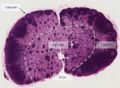
Structure


General
Grossly the lymph nodes are round or bean shaped and have an outer cortex and an inner medulla. Microscopically the nodes have follicles, paracortical zones and medullary cords and sinuses. At the hilum the medulla is present on the outer part of the node. Lymph nodes are located in series with lymphatic vessels. Afferent vessels enter the node on its convex side and efferent vessels exit on its concave side.
Capsule and reticular framework
The nodes are surrounded by a fibrous capsule that extends into the node as trabeculae, which provide an overall framework. Below the capsule is the subcapsular sinus. The nodes' parenchyma contain a fine network of reticular fibres and reticular cells. Reticular cells provide "scaffolding" for other cells as well as expressing surface complexes and substance to attract T cells, B cells and dendritic cells. The cortex has aggregations of B cells (in the follicles) in its outer region and a paracortex consisting of a rim of T cells surrounding these follicles. Dendritic cells are also found in close association with the T cells. The medulla contains medullary cords of cells (B cells, plasma cells and some macrophages) and between these cords is the medullary sinus lined with endothelial cells and macrophages.
Sinuses
Three sinuses are present:
- Subcapsular/cortical
- Where afferent vessels drain
- Trabecular
- Drain lymph from subcapsular to medullary sinuses
- Medullary
Antigens and transformed cells that pass through the sinuses are filtered by macrophages and removed from the lymph.
Follicles
Lymph nodes have two types of follicles, primary and secondary. Secondary follicles contain germinal centres (sites of B-cell proliferation) and have three layers.
- The central dark zone contains a high density of dividing centroblasts, B cells without surface Ig. These centroblasts migrate to the Basal light zone.
- In the basal light zone the B cells express surface Ig and become exposed to the follicular dendritic cells.
- Here there is a high rate of apoptosis but surviving cells migrate to the apical light zone.
- Apical light zone (mantle zone) which contains cells which are destined to become B memory (lymphoblasts) or plasma cells (plasmablasts).
Follicles in the cortex of a stimulated node are larger and have a pale germinal centre. Activated B cells differentiate into plasma and memory cells. Plasma cells migrate to the medullary cords and produce immunoglobulins.
High Endothelial Venules
High endothelial venules (HEV) are composed of cuboidal/columnar epithelium and are the major route for lymphocytes to enter the lymph node. HEV contain a large number of aquaporin-1 channels allowing for a large uptake of water which in turn drives lymph flow through the cortex. This fluid is then returned directly to the bloodstream. The venules are the source of most of the node's T cells and B cells and express selectins (receptors for lymphocytes primed with antigens).
HEV express CD34 and GlyCAM-1 which bind to L-selectin on naive lymphocytes. This allows circulating lymphocytes to recognise when they have reached a secondary lymphoid organ stimulating them to leave the bloodstream and enter the lymphatic tissue.
Pig Lymph Node
As well as dolphins, hippopotamuses and rhinoceroses
The structure of the pig lymph node is inverted compared with that of most mammals.
- Most follicles are found deep in the paracortex
- The paracortex is surrounded by loose medullary tissue
- Afferent lymphatics enter at the hilus
- Connect with para-trabecular sinuses and exit from efferent lymphatics on the node surface.
- Blood vessels enter and leave at the hilus
Histology
Gross view
©Nottingham Uni 2008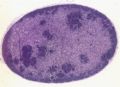
Gross view
©RVC 2008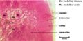
Gross view
©RVC 2008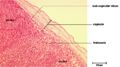
Capsule
©RVC 2008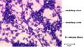
Medullary sinus
©RVC 2008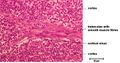
Trabecular sinus
©RVC 2008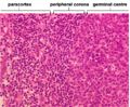
Follicular layers
©RVC 2008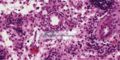
High endothelial venule
©Nottingham Uni 2008


Functions
The lymph nodes are secondary lymphoid tissue, and as the spleen removes antigens from the blood, lymph nodes remove antigens from tissue/lymph. Antigen presenting cells (B cells and T cells) migrate from peripheral tissue via afferent lymphatic vessels to the lymph nodes where they present their antigen to lymphocytes. B cells and T cells enter via the high endothelial venules by diapedesis and B cells migrate to the cortex while T cells to the deep cortex.
Antibodies and immunologically competent cells leave the lymph nodes via the efferent lymphatics.
| Lymph Nodes - Anatomy & Physiology Learning Resources | |
|---|---|
Anatomy Museum Resources |
Bovine Lymph Node Histology Bovine Lymph Node Histology - Germinal Centre |
| Originally funded by the RVC Jim Bee Award 2007 |
Webinars
Failed to load RSS feed from https://www.thewebinarvet.com/internal-medicine/webinars/feed: Error parsing XML for RSS