Oral Cavity Histology PowerPoint tutorial (1 of 2)
Click here to access the resource.
Resource Information
| Description
|
Oral Cavity Histology resource
PowerPoint
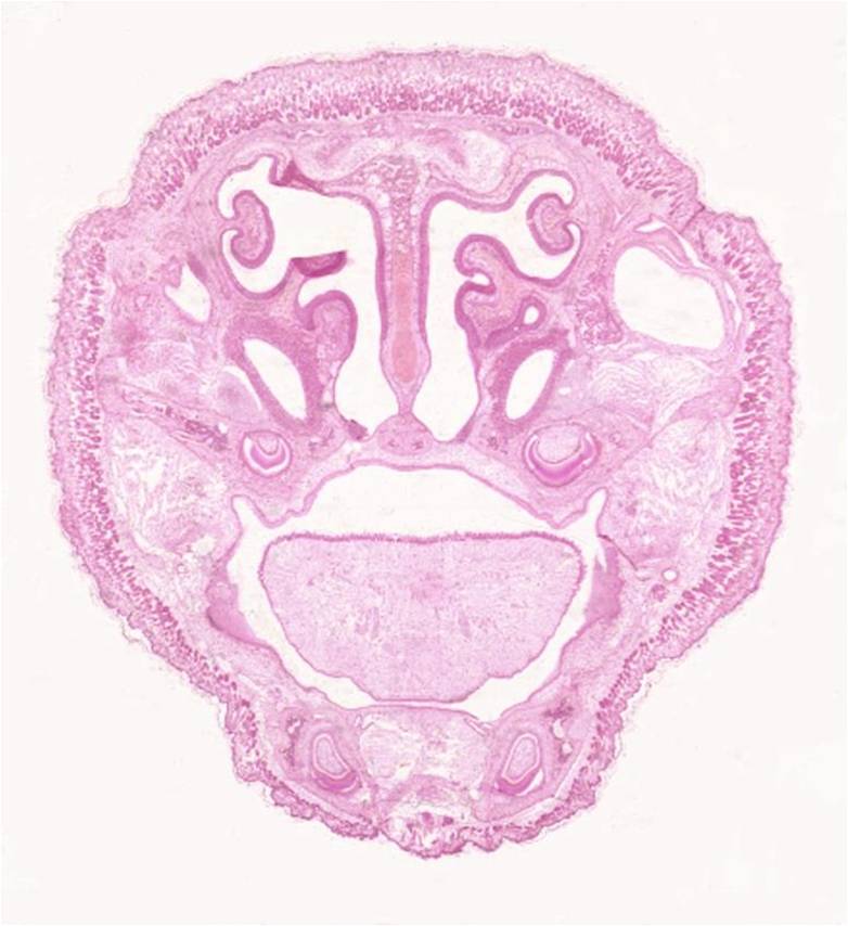
This is a tutorial on histology of the oral cavity. The PowerPoint contains many histological images of sections of the head, as well as images of different epithelial types and anatomy of the region, with the opportunity for self-assessment. This also features an easily accessible menu slide, allowing rapid navigation.
This is the first of 2 oral cavity tutorials
Duration = 41 slides
|
| Date
|
2011
|
| Source
|
Royal Veterinary College
|
| Author
|
John Bredl
|
| Licensing
|
|
Bone and Cartilage Histology PowerPoint tutorial (2 of 2) - Developmental
Click here to access the resource.
Resource Information
| Description
|
Oral Cavity Histology resource
PowerPoint
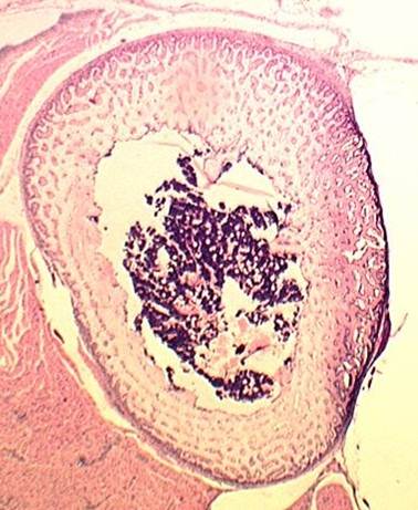
This is a tutorial on Bone and Cartilage Histology. The PowerPoint contains many histological images of different bone and cartilage cells and types with a focus on developmental bone and cartilage histology. This also features an easily accessible menu slide, allowing rapid navigation.
This is the second of 2 bone and cartilage tutorials
Duration = 35 slides
|
| Date
|
2011
|
| Source
|
Royal Veterinary College
|
| Author
|
John Bredl
|
| Licensing
|
|