Difference between revisions of "Parathyroid Glands - Anatomy & Physiology"
| Line 3: | Line 3: | ||
The parathyroid glands are multiple (generally four) small glands, approximately 1-2mm in length are located about the cranial trachea. Generally, there are two internal glands embedded within the [[Thyroid Gland - Anatomy & Physiology|thyroid Glands]], and two external glands are outside the thyroid tissue. However, all of the parathyroid tissue may be embedded within the thyroid gland itself. In the Horse, there are 'nests' of parathyroid tissue along the neck to the thoracic inlet. | The parathyroid glands are multiple (generally four) small glands, approximately 1-2mm in length are located about the cranial trachea. Generally, there are two internal glands embedded within the [[Thyroid Gland - Anatomy & Physiology|thyroid Glands]], and two external glands are outside the thyroid tissue. However, all of the parathyroid tissue may be embedded within the thyroid gland itself. In the Horse, there are 'nests' of parathyroid tissue along the neck to the thoracic inlet. | ||
| − | + | ==Embryology== | |
The parathyroid glands originate from the endoderm of pharyngeal pouches III and IV. The internal gland from pouch IV and the external from pouch III. | The parathyroid glands originate from the endoderm of pharyngeal pouches III and IV. The internal gland from pouch IV and the external from pouch III. | ||
Revision as of 17:10, 6 January 2011
Anatomy
The parathyroid glands are multiple (generally four) small glands, approximately 1-2mm in length are located about the cranial trachea. Generally, there are two internal glands embedded within the thyroid Glands, and two external glands are outside the thyroid tissue. However, all of the parathyroid tissue may be embedded within the thyroid gland itself. In the Horse, there are 'nests' of parathyroid tissue along the neck to the thoracic inlet.
Embryology
The parathyroid glands originate from the endoderm of pharyngeal pouches III and IV. The internal gland from pouch IV and the external from pouch III.
Histology
The parathyroids are histologically easy to distinguish from the thyroid.

The thyroid has a characteristic follicular structure, whereas the the parathyroid consists of densely packed cells, of two types:
1. Chief cells (Principal Cells)
These are the predominant cell type. They stain darker when they are active and are smaller than oxyphil cells. They manufacture Parathyroid Hormone (PTH).
2. Oxyphil cells
These fewer in number than chief cells. They stain lighter and are larger than chief cells. They have an unknown function.
Grossly, the parathyroids are difficult to differentiate from thyroid tissue or fat. A parathyroid gland may be accidentally removed during thyroidectomy. Care must therefore be taken if the second thryoid is removed to leave the parathyroid intact, otherwise hypoparathyroidism may ensue.
Histology Gallery
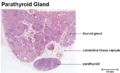
Parathyroid Gland Low Power 1
©RVC 2008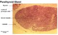
Parathyroid Gland Low Power 2
©RVC 2008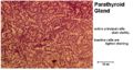
Parathyroid Gland with active Principal Cells
©RVC 2008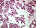
Normal Parathyroid Gland
Image courtesy of of Biomed Image Archive.


Blood Supply and Innervation
| Arteries | Veins | Nerve | Precursor |
|---|---|---|---|
| Superior thyroid artery | Superior thyroid vein | Middle cervical ganglion | Neural crest mesenchyme |
| Inferior thyroid artery | Middle thyroid vein | Inferior cervical ganglion | 3rd and 4th pharyngeal pouch endoderm |
| .</white> | Inferior thyroid vein | .</white> | .</white> |
Physiology
The sole function of the parathyroid gland is to maintain Calcium Homeostasis. Calcium homeostasis is, amongst other things, important for maintaining the function of the nervous and muscular systems. When blood calcium levels drop below a certain point, calcium-sensing receptors in the parathyroid gland are activated to release hormone into the blood. The hormone produced by the parathyroid gland (Parathyroid Hormone) also has an effect on phosphorus homeostasis.
Links
Click here for the parathyroid glands flashcards.