Difference between revisions of "Uterus - Anatomy & Physiology"
Fiorecastro (talk | contribs) |
|||
| (38 intermediate revisions by 7 users not shown) | |||
| Line 1: | Line 1: | ||
| − | {{ | + | {{OpenPagesTop}} |
| − | |||
| − | |||
| − | |||
| − | |||
| − | |||
| − | |||
| − | |||
| − | }} | ||
| − | |||
| − | |||
==Introduction== | ==Introduction== | ||
The Uterus is the organ of pregnancy as this is where implantation and development of the feotus occurs. The Uterus is the reproductive organ with the most species variations. These variations occur in both the anatomical types of uterus as well as the uterine horn appearance and endometrial linings. | The Uterus is the organ of pregnancy as this is where implantation and development of the feotus occurs. The Uterus is the reproductive organ with the most species variations. These variations occur in both the anatomical types of uterus as well as the uterine horn appearance and endometrial linings. | ||
| Line 17: | Line 7: | ||
'''Duplex Uteri''' | '''Duplex Uteri''' | ||
*Completely separate uterine horns each with their own cervical canal. Duplex uteri are seen in rabbits and marsupials. | *Completely separate uterine horns each with their own cervical canal. Duplex uteri are seen in rabbits and marsupials. | ||
| − | *As well as having 2 uterine horns and cervical canals | + | *As well as having 2 uterine horns and cervical canals marsupials also have two vaginas. |
'''Bicornuate Uteri''' | '''Bicornuate Uteri''' | ||
*(Previously Bipartite Uteri) Bicornuate uteri have a relatively small uterine body and two uterine horns of varying development. | *(Previously Bipartite Uteri) Bicornuate uteri have a relatively small uterine body and two uterine horns of varying development. | ||
| − | *A | + | *A relatively large uterine body and short, poorly developed uterine horns, as seen in the mare, are due to a high degree of paramesonephric duct fusion. |
| − | *Moderately developed uterine horns as in the | + | *Moderately developed uterine horns as in the cow, ewe and goat arise due to an intermediate degree of fusion. |
| − | *Long uterine horns and a small uterine body as seen in the sow, | + | *Long uterine horns and a small uterine body as seen in the sow, bitch and queen arise due to a low degree of fusion of the paramesonephric ducts. |
| − | *This varying degree of paramesonephric duct fusion reflects the species differences in litter sizes, | + | *This varying degree of paramesonephric duct fusion reflects the species differences in litter sizes, monotocous species having short horns and polytocous animals having much longer uterine horns. |
'''Simplex Uteri''' | '''Simplex Uteri''' | ||
*Complete fusion of the paramesonephric ducts forming a single uterine body with no uterine horns. This is seen in primates and especially humans. | *Complete fusion of the paramesonephric ducts forming a single uterine body with no uterine horns. This is seen in primates and especially humans. | ||
==Uterine Horn Appearance== | ==Uterine Horn Appearance== | ||
| + | [[Image:Mare Uterus.jpg|thumb|right|200px|The Mare Uterus- Courtesy of A.Crook, Copyright RVC 2008]] | ||
| + | [[Image:Dog Uteri.jpg|thumb|right|200px|Radiograph showing the Divergent Uterine Horns of the Bitch- Courtesy of A.Crook, Copyright RVC 2008]] | ||
{| style="width:75%; height:200px" border="1" | {| style="width:75%; height:200px" border="1" | ||
| Line 48: | Line 40: | ||
|- | |- | ||
|} | |} | ||
| + | |||
==Structure== | ==Structure== | ||
| − | + | ||
=== Perimetrium === | === Perimetrium === | ||
| − | *Serosal connective tissue layer continuous with the [[ | + | *Serosal connective tissue layer continuous with the [[Broad Ligament - Anatomy & Physiology|Broad Ligament]]. |
=== Myometrium === | === Myometrium === | ||
| Line 77: | Line 70: | ||
==Anatomical Location & Boundaries== | ==Anatomical Location & Boundaries== | ||
*The Uterus body and Uterine horns are located within the abdominal cavity dorsal to the Intestinal mass. | *The Uterus body and Uterine horns are located within the abdominal cavity dorsal to the Intestinal mass. | ||
| − | *The uterus is attached to the dorsal body wall via the Mesometrium [[ | + | *The uterus is attached to the dorsal body wall via the Mesometrium [[Broad Ligament - Anatomy & Physiology|Broad Ligament]]. |
==Histology== | ==Histology== | ||
The appearance of the uterus varies with the stages of the oestrus cycle. | The appearance of the uterus varies with the stages of the oestrus cycle. | ||
| − | + | <center><gallery widths=150px> | |
| + | Image:Uterus Histology.jpg|Histological Section of the Feline Uterus during Oestrus- Courtesy of J.Bredl, Copyright RVC 2008 | ||
| + | Image:Uterus Oestrus.jpg|Histological Section of the Feline Uterus during Oestrus- Courtesy of J.Bredl, Copyright RVC 2008 | ||
| + | Image:Uterus Luteal Phase.jpg|Histological Section of the Feline Uterus during the Luteal Phase- Courtesy of J.Bredl, Copyright RVC 2008 | ||
| + | Image:Uterus Anoestrus.jpg|Histological Section of the Canine Uterus during Anoestrus- Courtesy of J.Bredl, Copyright RVC 2008 | ||
| + | </gallery></center> | ||
=== Follicular phase-Oestrus === | === Follicular phase-Oestrus === | ||
| − | |||
*Increasing numbers of uterine glands developing and elongating within the endometrium due to the influence of Oestrogens produced by the developing follicles. | *Increasing numbers of uterine glands developing and elongating within the endometrium due to the influence of Oestrogens produced by the developing follicles. | ||
*General increase in the thickness of the endometrium. | *General increase in the thickness of the endometrium. | ||
*The epithelium lining the glands is Simple Columna epithelium. | *The epithelium lining the glands is Simple Columna epithelium. | ||
| + | === Luteal phase === | ||
| − | |||
| − | |||
| − | |||
| − | |||
| − | |||
| − | |||
| − | |||
| − | |||
| − | |||
| − | |||
| − | |||
| − | |||
| − | |||
| − | |||
| − | |||
*The endometrium is at its maximum thickness with a large number of highly developed glands. | *The endometrium is at its maximum thickness with a large number of highly developed glands. | ||
*This is the secretory phase for the endometrium. | *This is the secretory phase for the endometrium. | ||
| − | + | === Anoestrus === | |
| − | + | * The endothelium is relatively thin with little endometrial proliferation or gland development. | |
| − | + | ** This is due to the absence of ovarian steroids oestradiol and progesterone. | |
| − | *The endothelium is relatively thin with little endometrial proliferation or gland development. | + | * The glands are lined by Simple Cuboidal epithelium. |
| − | *The glands are lined by Simple Cuboidal epithelium. | ||
| − | |||
==Innervation== | ==Innervation== | ||
| − | The uterus is innervated by both | + | The uterus is innervated by both Sympathetic and Parasympathetic fibres which play a part in the regulation of uterine activity. This is highlighted by iatrogenic manipulation of parturition using β-Adrenoreceptor agonists for delaying parturition and antagonists for inducing parturition. However, uterine activity and normal parturition can be acheived when these nerves are severed. |
==Vasculature== | ==Vasculature== | ||
*Uterine branch of the Ovarian artery supplies the cranial parts of the Uterine horns. | *Uterine branch of the Ovarian artery supplies the cranial parts of the Uterine horns. | ||
| − | *Uterine artery supplies the rest of the uterine horns and the uterine body. This is a branch off the Internal Iliac artery in most domestic species, except the Mare where | + | *Uterine artery supplies the rest of the uterine horns and the uterine body. This is a branch off the Internal Iliac artery in most domestic species, except the Mare where instead it is a branch off the External Iliac artery. The Uterine artery and the Ovarian artery anastomose within the Broad ligament. |
| + | |||
| + | {{Learning | ||
| + | |dragster =[[Comparative Female Reproductive Anatomy resource]] | ||
| + | |videos = [[Video: Bovine Pregnant Uterus|Bovine pregnant uterus potcast]]<br>[[Video: Porcine uterus potcast|Porcine uterus potcast]]<br>[[Video: Bovine pregnant uterus potcast 2|Bovine pregnant uterus potcast 2]]<br>[[Video: Lateral view of the pelvic cavity and reproductive tract of the cow potcast|Lateral view of the pelvic cavity and reproductive tract of the cow potcast]]<br>[[Video: Female reproductive tract dissection|Female reproductive tract dissection]]<br>[[Video: Dissected equine abdomen showing ovary and uterus|Dissected equine abdomen showing ovary and uterus]]<br>[[Video: Female genital tract from a pregnant mare dissection|Female genital tract from a pregnant mare dissection]]<br>[[Video: Mare ovary and uterus dissection|Mare ovary and uterus dissection]]<br>[[Video: Isolated mare uterus potcast|Isolated mare uterus potcast]]<br>[[Video: Ewe uterine horns potcast|Ewe uterine horns potcast]]<br>[[Video: Ewe uterine horns potcast|Ewe uterine horns potcast]]<br>[[Video: Ovine uterus in late pregnancy dissection|Ovine uterus in late pregnancy dissection]]<br>[[Video: Ovine uterus and associated structures dissection|Ovine uterus and associated structures dissection]]<br>[[Video: Porcine uterus potcast|Porcine uterus potcast]]<br>[[Video: Isolated porcine uterus dissection|Isolated porcine uterus dissection]]<br>[[Video: Dissected porcine uterus in situ|Dissected porcine uterus in situ]] | ||
| + | |powerpoints = [[Female Reproductive Tract Histology resource|Histology of the female reproductive tract]] | ||
| + | }} | ||
| − | == | + | ==Webinars== |
| + | <rss max="10" highlight="none">https://www.thewebinarvet.com/urogenital-and-reproduction/webinars/feed</rss> | ||
| + | [[Category:Female Reproduction]] | ||
| + | [[Category:Bullet Points]] | ||
Latest revision as of 17:30, 7 November 2022
Introduction
The Uterus is the organ of pregnancy as this is where implantation and development of the feotus occurs. The Uterus is the reproductive organ with the most species variations. These variations occur in both the anatomical types of uterus as well as the uterine horn appearance and endometrial linings.
Anatomical Types of Uteri
Duplex Uteri
- Completely separate uterine horns each with their own cervical canal. Duplex uteri are seen in rabbits and marsupials.
- As well as having 2 uterine horns and cervical canals marsupials also have two vaginas.
Bicornuate Uteri
- (Previously Bipartite Uteri) Bicornuate uteri have a relatively small uterine body and two uterine horns of varying development.
- A relatively large uterine body and short, poorly developed uterine horns, as seen in the mare, are due to a high degree of paramesonephric duct fusion.
- Moderately developed uterine horns as in the cow, ewe and goat arise due to an intermediate degree of fusion.
- Long uterine horns and a small uterine body as seen in the sow, bitch and queen arise due to a low degree of fusion of the paramesonephric ducts.
- This varying degree of paramesonephric duct fusion reflects the species differences in litter sizes, monotocous species having short horns and polytocous animals having much longer uterine horns.
Simplex Uteri
- Complete fusion of the paramesonephric ducts forming a single uterine body with no uterine horns. This is seen in primates and especially humans.
Uterine Horn Appearance


| Species | Mare | Cow | Ewe | Sow | Bitch | Queen |
|---|---|---|---|---|---|---|
| Uterine Horn Appearance | Straight and divergent | Round | Round | Intestinal loop appearance | Straight and divergent | Straight |
Structure
Perimetrium
- Serosal connective tissue layer continuous with the Broad Ligament.
Myometrium
- Muscularis layer made up from thick inner circular smooth muscle layer and thin outer longitudinal muscle layer.
- The circular and longitudinal layers are seperated by the Stratum Vasculare, a connective tissue layer containing the blood vessels and nerves of the uterine wall.
Endometrium
- Mucosa & Submucosa layer containing endometrial glands.
- The surface of the endometrium differs between species:
- Ruminants - Numerous round button like elevations of the endometrium called Caruncles occur. These are the sites of attachment and maternal tissue contribution to the placenta. Maternal Caruncle + feotal cotyledon = A placentome.
- Sow and Mare - uterine folds are present.
- Not shed in domestic species, only shed in primates.
Function
- Provides a suitable environment for embryo development and attachment. The secretions produced by the endometrial glands are important for maintaining the preimplantion embryo.
- In response to increasing amounts of oxytocin production by the corpus luteum during the luteal phase the endometrium produces luteolytic PGF2a to cause degeneration of the corpus luteum if the female is not pregnant.
- The uterus contributes varying amounts of maternal tissues towards the placenta.
- The myometrium is involved with sperm transport through the uterus towards the oviduct.
- Contractions of the myometrium during parturition is important for feotus and placenta expulsion.
Anatomical Location & Boundaries
- The Uterus body and Uterine horns are located within the abdominal cavity dorsal to the Intestinal mass.
- The uterus is attached to the dorsal body wall via the Mesometrium Broad Ligament.
Histology
The appearance of the uterus varies with the stages of the oestrus cycle.
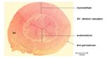
Histological Section of the Feline Uterus during Oestrus- Courtesy of J.Bredl, Copyright RVC 2008
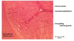
Histological Section of the Feline Uterus during Oestrus- Courtesy of J.Bredl, Copyright RVC 2008
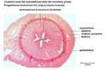
Histological Section of the Feline Uterus during the Luteal Phase- Courtesy of J.Bredl, Copyright RVC 2008
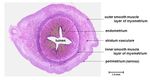
Histological Section of the Canine Uterus during Anoestrus- Courtesy of J.Bredl, Copyright RVC 2008


Follicular phase-Oestrus
- Increasing numbers of uterine glands developing and elongating within the endometrium due to the influence of Oestrogens produced by the developing follicles.
- General increase in the thickness of the endometrium.
- The epithelium lining the glands is Simple Columna epithelium.
Luteal phase
- The endometrium is at its maximum thickness with a large number of highly developed glands.
- This is the secretory phase for the endometrium.
Anoestrus
- The endothelium is relatively thin with little endometrial proliferation or gland development.
- This is due to the absence of ovarian steroids oestradiol and progesterone.
- The glands are lined by Simple Cuboidal epithelium.
Innervation
The uterus is innervated by both Sympathetic and Parasympathetic fibres which play a part in the regulation of uterine activity. This is highlighted by iatrogenic manipulation of parturition using β-Adrenoreceptor agonists for delaying parturition and antagonists for inducing parturition. However, uterine activity and normal parturition can be acheived when these nerves are severed.
Vasculature
- Uterine branch of the Ovarian artery supplies the cranial parts of the Uterine horns.
- Uterine artery supplies the rest of the uterine horns and the uterine body. This is a branch off the Internal Iliac artery in most domestic species, except the Mare where instead it is a branch off the External Iliac artery. The Uterine artery and the Ovarian artery anastomose within the Broad ligament.
| Uterus - Anatomy & Physiology Learning Resources | |
|---|---|
Test your knowledge using drag and drop boxes |
Comparative Female Reproductive Anatomy resource |
 Selection of relevant videos |
Bovine pregnant uterus potcast Porcine uterus potcast Bovine pregnant uterus potcast 2 Lateral view of the pelvic cavity and reproductive tract of the cow potcast Female reproductive tract dissection Dissected equine abdomen showing ovary and uterus Female genital tract from a pregnant mare dissection Mare ovary and uterus dissection Isolated mare uterus potcast Ewe uterine horns potcast Ewe uterine horns potcast Ovine uterus in late pregnancy dissection Ovine uterus and associated structures dissection Porcine uterus potcast Isolated porcine uterus dissection Dissected porcine uterus in situ |
 Selection of relevant PowerPoint tutorials |
Histology of the female reproductive tract |
Webinars
Failed to load RSS feed from https://www.thewebinarvet.com/urogenital-and-reproduction/webinars/feed: Error parsing XML for RSS