File:Braincosssection.jpg
Jump to navigation
Jump to search
No higher resolution available.
Braincosssection.jpg (622 × 478 pixels, file size: 31 KB, MIME type: image/jpeg)
Summary
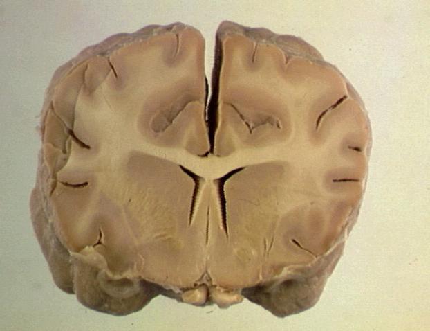
| Description |
Cross-section through the brain, showing the cerebrum, basal nuclei and lateral ventricle. The white and grey matter can be easily distinguished. |
|---|---|
| Date |
unknown |
| Source |
BioMed Image Archive |
| Author |
unknown |
| Permission (Reusing this file) |
See below |
Licensing
| This file is licensed under the Creative Commons Attribution Non-Commercial & No Derivative Works License |
File history
Click on a date/time to view the file as it appeared at that time.
| Date/Time | Thumbnail | Dimensions | User | Comment | |
|---|---|---|---|---|---|
| current | 13:31, 27 March 2008 | | 622 × 478 (31 KB) | Lizzies (talk | contribs) | Cross-section through the brain, showing the cerebrum, basal nuclei and lateral ventricle. The white and grey matter can be easily distinguished. |
You cannot overwrite this file.
File usage
The following 2 pages use this file: