Difference between revisions of "File:Hepatic stellate cells figure.jpg"
Jump to navigation
Jump to search
(uploaded a new version of "File:Hepatic stellate cells figure.jpg": {{Information |Description= Schematic presentation of hepatic stellate cells (HSC) located in the vicinity of adjacent hepatocytes (PC) beneath the sinusoidal endothelial ce) |
|||
| Line 1: | Line 1: | ||
| + | == Summary == | ||
| + | {{Information | ||
| + | |Description=Dog Dentition | ||
| + | |Source=Nottingham University | ||
| + | |Date=2008 | ||
| + | |Author=Unknown | ||
| + | |Permission=See below | ||
| + | }} | ||
| + | == Licensing: == | ||
| + | |||
| + | {{cc-att-2.0}} | ||
Revision as of 18:48, 19 January 2011
Summary
| Description |
Dog Dentition |
|---|---|
| Date |
2008 |
| Source |
Nottingham University |
| Author |
Unknown |
| Permission (Reusing this file) |
See below |
Licensing:
| This file is licensed under the Creative Commons Attribution Non-Commercial & No Derivative Works License |
File history
Click on a date/time to view the file as it appeared at that time.
| Date/Time | Thumbnail | Dimensions | User | Comment | |
|---|---|---|---|---|---|
| current | 18:31, 19 January 2011 | 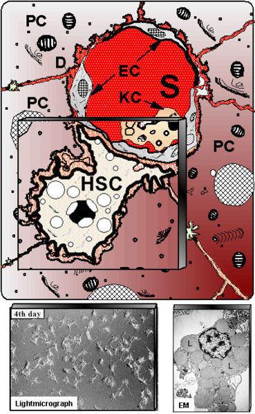 | 369 × 599 (85 KB) | Eca02csb (talk | contribs) | {{Information |Description= Schematic presentation of hepatic stellate cells (HSC) located in the vicinity of adjacent hepatocytes (PC) beneath the sinusoidal endothelial cells (EC). S – liver sinusoids; KC – Kupffer cells. Down left shows cultured HS |
You cannot overwrite this file.
File usage
The following page uses this file: