Difference between revisions of "Insulin"
m (Text replace - "Category:Alimentary System - Anatomy & Physiology" to "Alimentary System - Anatomy & Physiology") |
|||
| Line 27: | Line 27: | ||
| − | [[ | + | [[Alimentary System - Anatomy & Physiology]] |
Revision as of 12:49, 13 November 2011
Insulin is produced in the beta cells of the pancreatic islets of Langerhans and is released into the circulation to act on specific cell-surface receptors. Its release is stimulated by rising blood glucose concentration and it is principally insulin which is responsible for the post-prandial gluconeogenesis observed in humans and dogs. Several hormones (including corticosteroids, progesterone, oestrogen, growth hormone, glucagon and catecholamines) have an antagonistic effect to insulin and cause the blood glucose concentration to increase. Interruptions at any stage in this pathway may produce the clinical syndrome of diabetes mellitus.
Amylin is a protein which is produced normally in the beta cells at the same rate as insulin and has synergistic effects on many aspects of metabolism. In situations where the synthesis of insulin is increased due to insulin resistance (see diabetes mellitus), amylin is also produced in excess and it then forms aggregates that are deposited in the pancreatic tissue.
- Insulin secretion is stimulated under conditions of hyperglycaemia and inhibited under conditions of hypoglycaemia.
- It has an anabolic effect on energy sources.
- It is a small polypeptide consisting of two chains; A and B; which are linked by disulphide bonds.
- It is synthesised inactively as a pre-pro-hormone which undergoes cleavage to produce the active form.
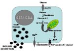
- Insulin is released when blood glucose levels are high. The process of insulin release occurs as follows:
- GLUT 2, which only operate at high glucose concentrations, are present in the plasma membranes of β cells.
- After a meal, glucose concentrations in the blood rise and glucose is taken into the cell.
- Glucose is metabolised inside the β cell which produces ATP.
- ATP inhibits potassium channels which depolarises the plasma membrane.
- Depolarisation causes calcium signalling, which stimulates insulin to be released.
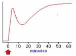
- Pre-formed granules of insulin are release initially, but this supply soon diminishes. This produces a decrease in plasma insulin levels. Insulin levels are restored as de novo insulin is synthesised and secreted.
- Insulin exerts it's anabolic effect on the liver, adipocytes and skeletal muscle.
- It binds to receptors on the cell membrane, which triggers an intracellular signalling cascade.
- This causes preformed granules containing GLUT 4 receptors to be transported and expressed at the cell membrane.
- Cells can now take up glucose from the blood and store it as glycogen or fat.
- Increased glucose levels inside the cell increases the rate of glycogenesis and an increase in the rate of production of triacylglycerides.
- There is an increase in basal metabolic rate in other tissues.
- Insulin regulates the reaction in glycolysis where PEP is converted into pyruvate. This reaction is regulated by the enzyme pyruvate kinase. Insulin activates this enzyme by dephosphorylation via protein phosphatase 1 (PP1). This increases metabolism.

