|
|
| (8 intermediate revisions by one other user not shown) |
| Line 4: |
Line 4: |
| | | | |
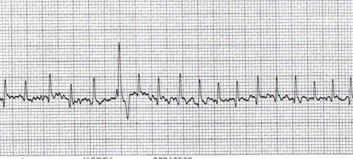
| | Mikey was admitted for an electrocardiogram, echocardiography and thoracic radiographs. The ECG is shown below: | | Mikey was admitted for an electrocardiogram, echocardiography and thoracic radiographs. The ECG is shown below: |
| − |
| |
| − |
| |
| − | ECG recording in lead II 25mm/sec 10mm/mV
| |
| | | | |
| | [[Image:Clinical Case 5 02.jpg|thumb|center|500px|(Courtesy of C. Antonczyk)]] | | [[Image:Clinical Case 5 02.jpg|thumb|center|500px|(Courtesy of C. Antonczyk)]] |
| | | | |
| | | | |
| − | [[Image:Clinical Case 5 03.jpg|thumb|center|500px|(Courtesy of C. Antonczyk)]]
| + | *Click [[Clinical Case 5 - Page 3|here]] to see a close up image under the x10 objective. |
| − | | |
| − | | |
| − | | |
| − | In order to reveal an answer, highlight the underlined or bulleted area using your mouse.
| |
| − | The number of bullet points doesn't necessarily indicate a strict number of answers.
| |
| − | | |
| − | | |
| − | For what reason might you perform an ECG?
| |
| − | *<font color="white"> In order to diagnose a rhythm disturbance. </font>
| |
| − | | |
| − | What is the average heart rate on the first half of this ECG?
| |
| − | *<font color="white"> This recording is 25mm/sec, so 25 of the small boxes represent a second and 5 of the larger boxes represent a second. If you count the number of beats starting from the first thick line over the following 4 seconds (20 of the larger squares) you get 15 beats. So in 4 seconds there were 15 beats. Hence in one minute there will be 15x15 beats. The average heart rate is 225 beats per minute. </font> | |
| − | | |
| − | Can you see any P waves? Why do you think this might be?
| |
| − | *<font color="white"> No. This dog has atrial fibrillation. P waves are absent. </font>
| |
| − | | |
| − | On the first half of the ECG, why does the 6th complex look different to the rest?
| |
| − | *<font color="white"> This complex is wider than the others indicating that it is of ventricular origin. It is a ventricular premature complex (VPC)</font>
| |
| − | | |
| − | On the second part of the ECG, what do we call the series of abnormal complexes?
| |
| − | *<font color="white"> This is a run of VPCs. As there are more than three in succession, this is called ventricular tachycardia. </font>
| |
| − | | |
| − | | |
| − | Click [[Clinical Case 5 - Page 3|here]] to see the results of the ultrasound. | |
| − | | |
| − | | |
| − | [[Category:Lost]]
| |
